Department of Physics, Korea Advanced Institute of Science and Technology (KAIST), 291 Daehak-ro, Yuseong-Gu, Daejeon, 34141, Republic of Korea.
KAIST Institute for Health Science and Technology, KAIST, Daejeon, 34141, Republic of Korea.
Sci Rep. 2018 Jun 15;8(1):9183. doi: 10.1038/s41598-018-27399-w.
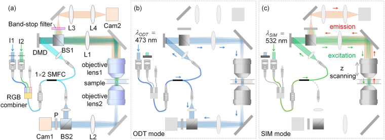
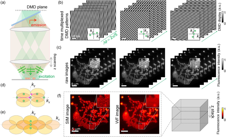
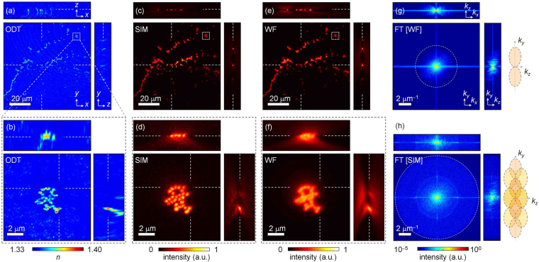
We present a multimodal approach for measuring the three-dimensional (3D) refractive index (RI) and fluorescence distributions of live cells by combining optical diffraction tomography (ODT) and 3D structured illumination microscopy (SIM). A digital micromirror device is utilized to generate structured illumination patterns for both ODT and SIM, which enables fast and stable measurements. To verify its feasibility and applicability, the proposed method is used to measure the 3D RI distribution and 3D fluorescence image of various samples, including a cluster of fluorescent beads, and the time-lapse 3D RI dynamics of fluorescent beads inside a HeLa cell, from which the trajectory of the beads in the HeLa cell is analyzed using spatiotemporal correlations.
我们提出了一种结合光学衍射层析成像(ODT)和三维结构光照明显微镜(SIM)的多模态方法,用于测量活细胞的三维(3D)折射率(RI)和荧光分布。数字微镜器件用于为 ODT 和 SIM 生成结构照明图案,这使得快速和稳定的测量成为可能。为了验证其可行性和适用性,该方法用于测量各种样品的 3D RI 分布和 3D 荧光图像,包括荧光珠簇和 HeLa 细胞内荧光珠的 3D RI 动力学的时程变化,从中使用时空相关分析了 HeLa 细胞内珠的轨迹。