University of Maryland Medical Center and Sheppard Pratt Health System, Department of Psychiatry, Baltimore, MD, USA.
Biomarker Discovery Center, New Jersey Institute for Successful Aging, Rowan University School of Osteopathic Medicine, Stratford, NJ, USA.
J Alzheimers Dis. 2020;74(1):345-361. doi: 10.3233/JAD-190962.
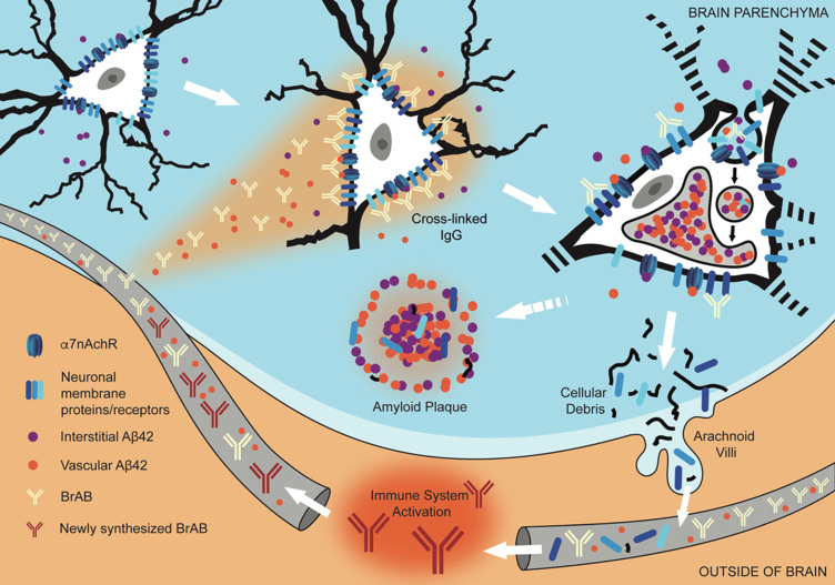
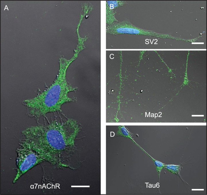
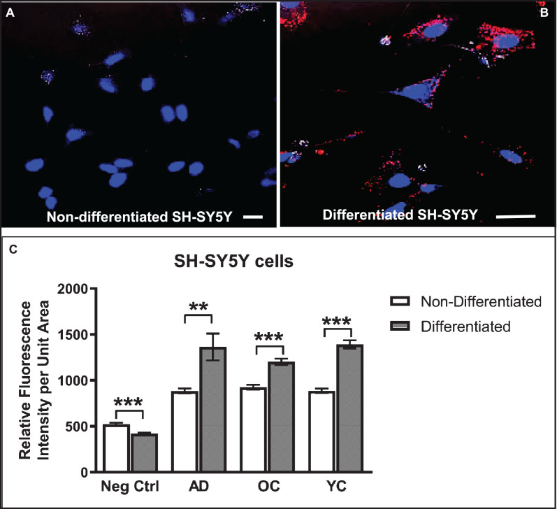
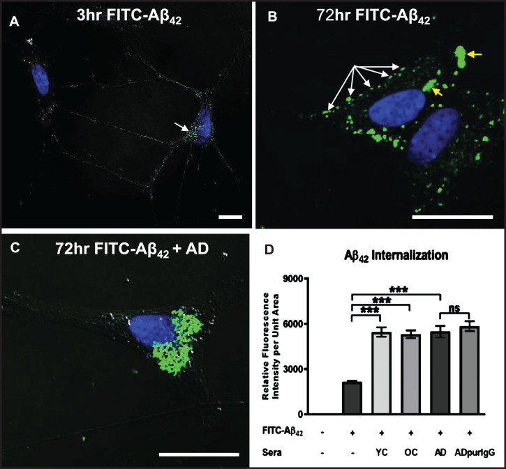
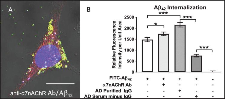
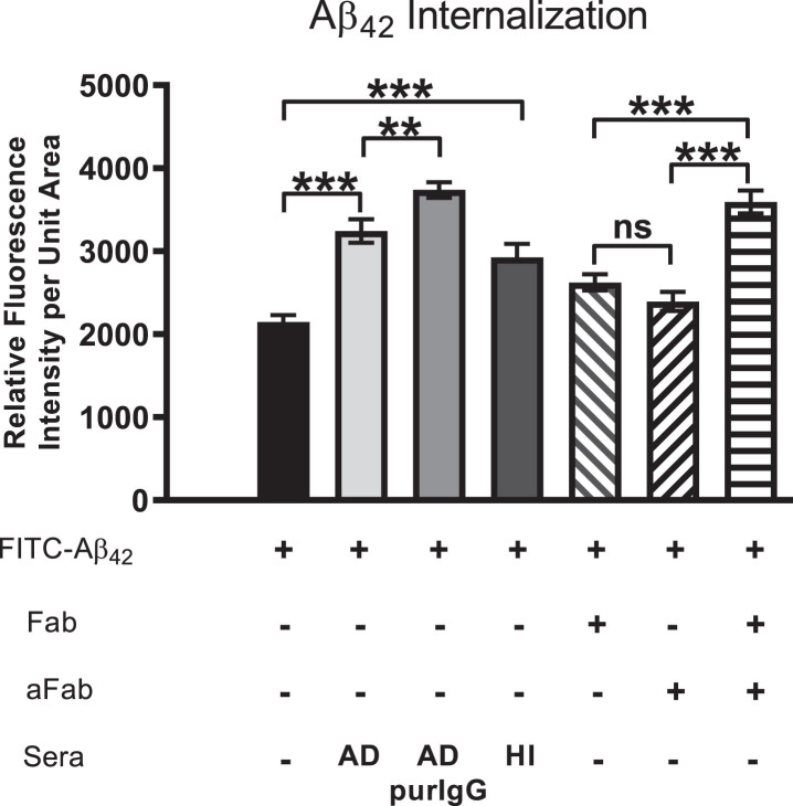
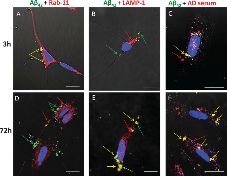
Blood-brain barrier (BBB) permeability is a recognized early feature of Alzheimer's disease (AD). In the present study, we examined consequences of increased BBB permeability on the development of AD-related pathology by tracking selected leaked plasma components and their interactions with neurons in vivo and in vitro. Histological sections of cortical regions of postmortem AD brains were immunostained to determine the distribution of amyloid-β1-42 (Aβ42), cathepsin D, IgG, GluR2/3, and alpha7 nicotinic acetylcholine receptor (α7nAChR). Results revealed that chronic IgG binding to pyramidal neurons coincided with internalization of Aβ42, IgG, GluR2/3, and α7nAChR as well as lysosomal compartment expansion in these cells in regions of AD pathology. To test possible mechanistic interrelationships of these phenomena, we exposed differentiated SH-SY5Y neuroblastoma cells to exogenous, soluble Aβ42 peptide and serum from AD and control subjects. The rate and extent of Aβ42 internalization in these cells was enhanced by serum containing neuron-binding IgG autoantibodies. This was confirmed by treating cells with individual antibodies specific for α7nAChR, purified IgG from AD or non-AD sera, and sera devoid of IgG, in the presence of 100 nM Aβ42. Initial co-localization of IgG, α7nAChR, and Aβ42 was temporally and spatially linked to early endosomes (Rab11) and later to lysosomes (LAMP-1). Aβ42 internalization was attenuated by treatment with monovalent F(ab) antibody fragments generated from purified IgG from AD serum and then rescued by coupling F(ab) fragments with divalent human anti-Fab. Overall, results suggest that cross-linking of neuron-binding autoantibodies targeting cell surface proteins can accelerate intraneuronal Aβ42 deposition in AD.
血脑屏障(BBB)通透性是阿尔茨海默病(AD)的一个公认的早期特征。在本研究中,我们通过体内和体外跟踪选定的渗漏血浆成分及其与神经元的相互作用,研究了 BBB 通透性增加对 AD 相关病理学发展的影响。对 AD 脑死后皮质区域的组织学切片进行免疫染色,以确定淀粉样蛋白-β1-42(Aβ42)、组织蛋白酶 D、IgG、GluR2/3 和α7 烟碱型乙酰胆碱受体(α7nAChR)的分布。结果表明,慢性 IgG 与锥体神经元结合,同时伴随着 Aβ42、IgG、GluR2/3 和α7nAChR 的内化以及这些细胞中溶酶体区室的扩张,这些都发生在 AD 病变区域。为了测试这些现象之间可能存在的机制关系,我们将分化的 SH-SY5Y 神经母细胞瘤暴露于外源性、可溶性 Aβ42 肽和来自 AD 和对照受试者的血清中。在这些细胞中,含有神经元结合 IgG 自身抗体的血清增强了 Aβ42 的内化速度和程度。这通过用针对α7nAChR 的特异性单克隆抗体、来自 AD 或非 AD 血清的纯化 IgG 以及在存在 100 nM Aβ42 的情况下不含 IgG 的血清处理细胞得到了证实。IgG、α7nAChR 和 Aβ42 的初始共定位在时间和空间上与早期内体(Rab11)相关,随后与溶酶体(LAMP-1)相关。用来自 AD 血清的纯化 IgG 产生的单价 F(ab')抗体片段处理后,Aβ42 的内化被减弱,然后用二价人抗 F(ab')片段将 F(ab')片段连接起来,使内化得到挽救。总的来说,结果表明,针对细胞表面蛋白的神经元结合自身抗体的交联可以加速 AD 中神经元内 Aβ42 的沉积。