• 文献检索
  • 文档翻译
  • 深度研究
  • 学术资讯
  • Suppr Zotero 插件Zotero 插件
  • 邀请有礼
  • 套餐&价格
  • 历史记录
应用&插件
Suppr Zotero 插件Zotero 插件浏览器插件Mac 客户端Windows 客户端微信小程序
定价
高级版会员购买积分包购买API积分包
服务
文献检索文档翻译深度研究API 文档MCP 服务
关于我们
关于 Suppr公司介绍联系我们用户协议隐私条款
关注我们

Suppr 超能文献

核心技术专利:CN118964589B侵权必究
粤ICP备2023148730 号-1Suppr @ 2026

文献检索

告别复杂PubMed语法,用中文像聊天一样搜索,搜遍4000万医学文献。AI智能推荐,让科研检索更轻松。

立即免费搜索

文件翻译

保留排版,准确专业,支持PDF/Word/PPT等文件格式,支持 12+语言互译。

免费翻译文档

深度研究

AI帮你快速写综述,25分钟生成高质量综述,智能提取关键信息,辅助科研写作。

立即免费体验

组织病理学证实的纤维结构不良患者的磁共振成像特征:系统评价。

Magnetic resonance imaging characteristics in patients with histopathologically proven fibrous dysplasia-a systematic review.

机构信息

Institute of Clinical Medicine, University of Eastern Finland, Yliopistonranta 1, 70210, Kuopio, Finland.

Department of Clinical Pathology, Kuopio University Hospital, Kuopio, Finland.

出版信息

Skeletal Radiol. 2020 Jun;49(6):837-845. doi: 10.1007/s00256-020-03388-x. Epub 2020 Feb 10.

DOI:10.1007/s00256-020-03388-x
PMID:32040603
原文链接:https://pmc.ncbi.nlm.nih.gov/articles/PMC7170814/
Abstract

OBJECTIVE

To examine the demographics, lesion location, and characteristic magnetic resonance imaging (MRI) findings in patients with histopathologically proven fibrous dysplasia (FD).

MATERIALS AND METHODS

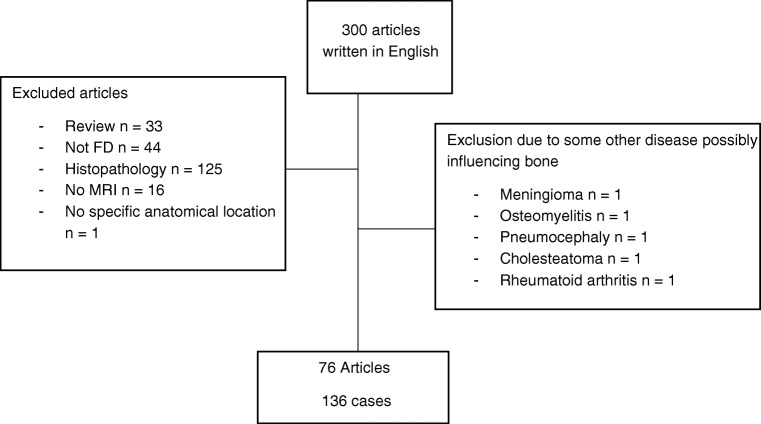
A systematic literature search of the MRI findings in patients with histologically proven FD was performed. Altogether, 76 articles with 136 patients were evaluated.

RESULTS

The mean age of the patients was 35.0 + - 18.5 years (range 1 month-75 years). Fifty-eight of the cases were females, 51 males, and in 27 gender was not defined. The most common locations were craniofacial (n = 55 (40%)), long bones (n = 31 (23%)), and spine (n = 24 (18%)). The monostotic form of FD was the most common. Signal intensities (SI) on T1-weighted images were predominantly hypointense (n = 46 (37%)). The SI was highly variable on T2-weighted images with hyperintensity being most common (n = 22 (18%)). Contrast enhancement was found in 75 (55%) FD patients. Secondary aneurysmal bone cysts (ABCs) and malignant transformation in patients without prior radiotherapy was found in some patients.

CONCLUSION

Current knowledge of the MRI findings in patients with FD is based mainly on case reports. SI in patients with FD is variable and contrast enhancement is common. FD may explain etiology of spinal bone tumor in some patients. FD with malignant transformation should be considered also in patients without prior radiotherapy. Further studies are needed to clarify if FD displays specific characteristics allowing it to be distinguished from other bone tumors.

摘要

目的

研究组织学证实的纤维结构不良(FD)患者的人口统计学、病变位置和特征性磁共振成像(MRI)表现。

材料与方法

对组织学证实 FD 患者的 MRI 表现进行系统的文献检索。共评估了 76 篇文章和 136 例患者。

结果

患者的平均年龄为 35.0±18.5 岁(1 个月-75 岁)。58 例为女性,51 例为男性,27 例未定义性别。最常见的部位为颅面(n=55(40%))、长骨(n=31(23%))和脊柱(n=24(18%))。FD 的单骨形式最常见。T1 加权图像的信号强度(SI)主要为低信号(n=46(37%))。T2 加权图像的 SI 高度可变,高信号最常见(n=22(18%))。75 例(55%)FD 患者有增强表现。一些患者出现继发性动脉瘤样骨囊肿(ABC)和未经放射治疗的恶性转化。

结论

目前对 FD 患者 MRI 表现的认识主要基于病例报告。FD 患者的 SI 是多变的,增强是常见的。FD 可能是一些患者脊柱骨肿瘤的病因。对于没有放射治疗史的患者,也应考虑 FD 发生恶性转化的可能性。需要进一步研究以明确 FD 是否具有可将其与其他骨肿瘤区分开来的特定特征。

https://cdn.ncbi.nlm.nih.gov/pmc/blobs/a26a/7170814/c2c5313813e6/256_2020_3388_Fig1_HTML.jpg

相似文献

1
Magnetic resonance imaging characteristics in patients with histopathologically proven fibrous dysplasia-a systematic review.组织病理学证实的纤维结构不良患者的磁共振成像特征:系统评价。
Skeletal Radiol. 2020 Jun;49(6):837-845. doi: 10.1007/s00256-020-03388-x. Epub 2020 Feb 10.
2
CT and MRI of fibrous dysplasia of the spine.脊柱纤维结构不良的 CT 和 MRI。
Br J Radiol. 2012 Jul;85(1015):996-1001. doi: 10.1259/bjr/81329736. Epub 2011 Dec 13.
3
Fibrous dysplasia: MR imaging characteristics with radiopathologic correlation.骨纤维异常增殖症:具有放射病理学相关性的磁共振成像特征
AJR Am J Roentgenol. 1996 Dec;167(6):1523-7. doi: 10.2214/ajr.167.6.8956590.
4
Unusual Presentation of Fibrous Dysplasia in an Elderly Patient.老年患者纤维发育不良的罕见表现
J Radiol Case Rep. 2019 Feb 28;13(2):26-33. doi: 10.3941/jrcr.v13i2.3379. eCollection 2019 Feb.
5
Milk cloud appearance-a characteristic sign of fibrous dysplasia on contrast-enhanced MR imaging.牛奶样外观——增强磁共振成像上纤维结构不良的特征性征象。
Eur Radiol. 2019 Jul;29(7):3424-3430. doi: 10.1007/s00330-019-06245-1. Epub 2019 May 6.
6
Malignant transformation in monostotic fibrous dysplasia: clinical features, imaging features, outcomes in 10 patients, and review.单骨型骨纤维异常增殖症的恶性转化:10例患者的临床特征、影像特征、治疗结果及文献复习
Medicine (Baltimore). 2015 Jan;94(3):e369. doi: 10.1097/MD.0000000000000369.
7
CT and MR Imaging in a Large Series of Patients with Craniofacial Fibrous Dysplasia.大量颅面骨纤维发育不良患者的CT和MR成像
Pol J Radiol. 2015 May 4;80:232-40. doi: 10.12659/PJR.893425. eCollection 2015.
8
Ground-glass pattern fibrous dysplasia of frontal sinus.额窦磨玻璃样纤维结构不良
JBR-BTR. 2013 Nov-Dec;96(6):378-80. doi: 10.5334/jbr-btr.467.
9
[Fibrous dysplasia: its appearance in the magnetic resonance tomogram].[骨纤维发育不良:其在磁共振断层扫描中的表现]
Rofo. 1989 Sep;151(3):338-41. doi: 10.1055/s-2008-1047189.
10
Secondary Aneurysmal Bone Cyst in a Craniofacial Fibrous Dysplasia: Case Report.颅面部骨纤维发育不良中的继发性动脉瘤样骨囊肿:病例报告
Brain Tumor Res Treat. 2018 Oct;6(2):86-91. doi: 10.14791/btrt.2018.6.e15.

引用本文的文献

1
Treatment approach with clinical follow-up in monostotic fibrous dysplasia: a case series.单发性骨纤维发育不良的临床随访治疗方法:病例系列
J Med Case Rep. 2025 May 13;19(1):223. doi: 10.1186/s13256-025-05261-8.
2
Fibrous Dysplasia in Humerus Shaft - Fixation or Curettage?: A Case Report.肱骨干纤维发育不良——固定还是刮除术?:一例报告
J Orthop Case Rep. 2025 May;15(5):50-55. doi: 10.13107/jocr.2025.v15.i05.5552.
3
Benign Notochordal Cell Tumours: Case Report and Literature Review.良性脊索细胞瘤:病例报告及文献综述

本文引用的文献

1
Malignant transformation in monostotic fibrous dysplasia: clinical features, imaging features, outcomes in 10 patients, and review.单骨型骨纤维异常增殖症的恶性转化:10例患者的临床特征、影像特征、治疗结果及文献复习
Medicine (Baltimore). 2015 Jan;94(3):e369. doi: 10.1097/MD.0000000000000369.
2
Locally aggressive fibrous dysplasia mimicking malignancy: a report of four cases and review of the literature.酷似恶性肿瘤的局部侵袭性骨纤维异常增殖症:4例报告并文献复习
Clin Orthop Relat Res. 2015 Feb;473(2):742-50. doi: 10.1007/s11999-014-3926-x. Epub 2014 Sep 12.
3
Fibrous dysplasia.
Diagnostics (Basel). 2024 Jun 23;14(13):1330. doi: 10.3390/diagnostics14131330.
4
Imaging approach to pediatric calvarial bulges.小儿颅骨膨出的影像学方法。
Pediatr Radiol. 2024 Sep;54(10):1603-1617. doi: 10.1007/s00247-024-05967-9. Epub 2024 Jun 28.
5
New technologies in oral radiology as a diagnostic aid for monostotic fibrous dysplasia: a review.口腔放射学中的新技术作为单骨型纤维发育不良的诊断辅助手段:综述
Rev Cient Odontol (Lima). 2021 Dec 9;9(4):e089. doi: 10.21142/2523-2754-0904-2021-089. eCollection 2021 Oct-Dec.
6
Multimodal imaging diagnosis for bone fibrous dysplasia malignant transformation: A case report.骨纤维发育不良恶变的多模态成像诊断:一例报告
Biomed Rep. 2023 Sep 1;19(4):73. doi: 10.3892/br.2023.1655. eCollection 2023 Oct.
7
Polyostotic Fibrous Dysplasia in a Six-year-Old Boy.一名六岁男孩的多骨型纤维发育不良
Mol Imaging Radionucl Ther. 2023 Jun 20;32(2):186-190. doi: 10.4274/mirt.galenos.2023.79095.
8
Comparison of MRI Findings among Osteofibrous Dysplasia, Fibrous Dysplasia, and NonOssifying Fibroma of the Long Bone.长骨骨纤维异常增殖症、骨纤维结构不良和非骨化性纤维瘤的MRI表现比较。
Indian J Radiol Imaging. 2023 Jan 13;33(2):150-156. doi: 10.1055/s-0042-1760363. eCollection 2023 Apr.
9
Benign incidental do-not-touch bone lesions.良性偶然勿触骨病变。
Br J Radiol. 2023 Feb;96(1142):20211334. doi: 10.1259/bjr.20211334. Epub 2022 May 31.
10
Successful treatment with denosumab for pelvic fibrous dysplasia: A case report and review of the literature.成功使用地舒单抗治疗骨盆纤维结构不良:病例报告及文献复习。
Medicine (Baltimore). 2021 Dec 10;100(49):e28138. doi: 10.1097/MD.0000000000028138.
纤维结构不良。
Arch Pathol Lab Med. 2013 Jan;137(1):134-8. doi: 10.5858/arpa.2012.0013-RS.
4
Endoscopic transphenoidal approach for fibrous dysplasia of clivus, tuberculum sellae and sphenoid sinus; report of three cases.内镜经蝶入路治疗斜坡、鞍结节及蝶窦骨纤维异常增殖症:三例报告
Turk Neurosurg. 2012;22(5):662-6. doi: 10.5137/1019-5149.JTN.4060-10.0.
5
Aneurysmal bone cyst concomitant with fibrous dysplasia in the frontal bone.额骨动脉瘤样骨囊肿合并骨纤维发育不良。
Neurosciences (Riyadh). 2010 Oct;15(4):280-3.
6
Maxillofacial fibrous dysplasia: personal experience with gadoliniumenhanced magnetic resonance imaging.颌面部纤维性发育异常:钆增强磁共振成像的个人经验
Radiol Med. 2008 Dec;113(8):1198-210. doi: 10.1007/s11547-008-0329-7. Epub 2008 Oct 4.
7
Fibrous dysplasia of bone and McCune-Albright syndrome.骨纤维发育不良和麦库恩-奥尔布赖特综合征
Best Pract Res Clin Rheumatol. 2008 Mar;22(1):55-69. doi: 10.1016/j.berh.2007.11.004.
8
Monostotic fibrous dysplasia of the middle phalanx.中节指骨单发性骨纤维异常增殖症
Hand Surg. 2007;12(2):91-5. doi: 10.1142/S0218810407003432.
9
Magnetic resonance imaging appearances of fibrous dysplasia.
Br J Radiol. 2005 Dec;78(936):1104-15. doi: 10.1259/bjr/73852511.
10
Fibrous dysplasia. Pathophysiology, evaluation, and treatment.骨纤维异常增殖症。病理生理学、评估与治疗。
J Bone Joint Surg Am. 2005 Aug;87(8):1848-64. doi: 10.2106/JBJS.D.02942.