Department of Surgery, Radboud university medical center, Nijmegen, The Netherlands.
Department of Radiology and Nuclear Medicine, Radboud university medical center, Nijmegen, The Netherlands.
Mol Diagn Ther. 2020 Apr;24(2):191-200. doi: 10.1007/s40291-020-00448-9.
Tumor-targeted imaging is a promising technique for the detection of lymph node metastases (LNM) and primary tumors. It remains unclear which biomarker is the most suitable target to distinguish malignant from healthy tissue in esophageal adenocarcinoma (EAC).
We performed an immunohistochemistry study to identify viable tumor markers for tumor-targeted imaging of EAC.
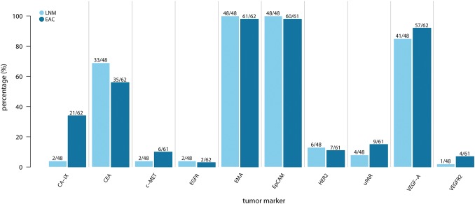
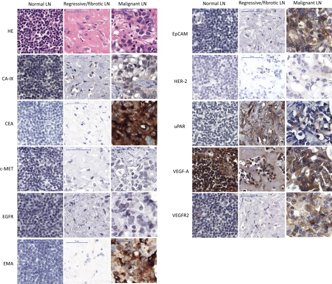
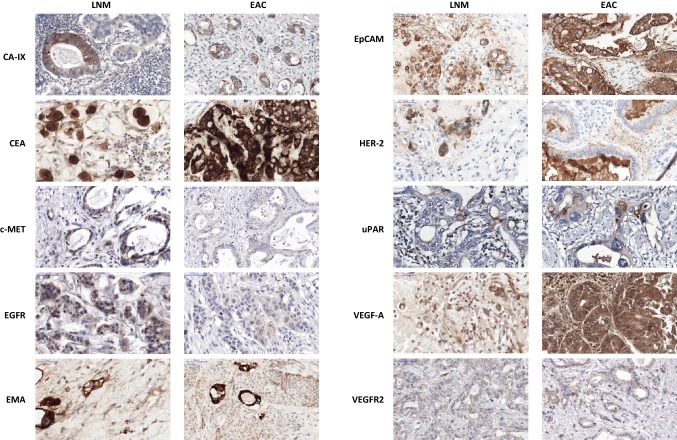
We used samples from 72 patients with EAC to determine the immunohistochemical expression of ten potential tumor biomarkers for EAC (carbonic anhydrase IX [CA-IX], carcinoembryonic antigen [CEA], hepatic growth factor receptor, epidermal growth factor receptor, epithelial membrane antigen [EMA], epithelial cell adhesion molecule [EpCAM], human epidermal growth factor receptor 2 [HER-2], urokinase plasminogen activator receptor, vascular endothelial growth factor-A [VEGF-A], and VEGF receptor 2). Immunohistochemistry was performed on tissue microarrays of LNM (n = 48), primary EACs (n = 62), fibrotic tissues (n = 11), nonmalignant lymph nodes (n = 24), and normal esophageal and gastric tissues (n = 40). Tumor marker staining was scored on intensity and percentage of positive cells.
EMA and EpCAM showed strong expression in LNM (> 95%) and primary EACs (> 95%). Significant expression was also observed for LNM and EAC using VEGF-A (85 and 92%), CEA (68 and 54%), and CA-IX (4 and 34%). The other tumor biomarkers showed expression of 0-15% for LNM and primary EAC. Except for VEGF-A, nonmalignant lymph node staining was scored as slight or absent.
High expression rates and correlation between LNM in EAC combined with low expression rates in healthy lymph nodes and esophagus tissues were observed for EpCAM and CEA, meaning these are promising targets for tumor-targeted imaging approaches for lymph nodes in patients with EAC.
肿瘤靶向成像技术是一种很有前途的方法,可用于检测淋巴结转移(LNM)和原发性肿瘤。目前尚不清楚哪种生物标志物最适合作为区分食管腺癌(EAC)中恶性组织和健康组织的靶标。
我们进行了免疫组织化学研究,以确定用于 EAC 肿瘤靶向成像的可行肿瘤标志物。
我们使用 72 例 EAC 患者的样本,确定了 10 种潜在的 EAC 肿瘤标志物(碳酸酐酶 IX [CA-IX]、癌胚抗原 [CEA]、肝细胞生长因子受体、表皮生长因子受体、上皮膜抗原 [EMA]、上皮细胞黏附分子 [EpCAM]、人表皮生长因子受体 2 [HER-2]、尿激酶型纤溶酶原激活物受体、血管内皮生长因子-A [VEGF-A]和 VEGF 受体 2)的免疫组织化学表达。对 LNM(n=48)、原发性 EAC(n=62)、纤维化组织(n=11)、非恶性淋巴结(n=24)和正常食管及胃组织(n=40)的组织微阵列进行免疫组织化学染色。根据阳性细胞的强度和百分比对肿瘤标志物染色进行评分。
EMA 和 EpCAM 在 LNM(>95%)和原发性 EAC(>95%)中表达强烈。VEGF-A(85%和 92%)、CEA(68%和 54%)和 CA-IX(4%和 34%)在 LNM 和 EAC 中也有显著表达。其他肿瘤标志物在 LNM 和原发性 EAC 中的表达率为 0-15%。除 VEGF-A 外,非恶性淋巴结染色评分较低或未见。
在 EAC 中,LNM 的高表达率和与健康淋巴结和食管组织的低表达率之间存在相关性,观察到 EpCAM 和 CEA 的表达率较高,这意味着它们是用于 EAC 患者淋巴结肿瘤靶向成像方法的有前途的靶标。