,.
Invest Ophthalmol Vis Sci. 2020 Feb 7;61(2):14. doi: 10.1167/iovs.61.2.14.
Experimental access to specific cell subtypes is essential for deciphering the complexity of retinal networks. Here, we characterized the selective labeling, caused by ectopic transgene expression, of two atypical retinal neurons in the ChAT-Channelrhodopsin-2 (ChR2)-EYFP mouse.
Retinal sections and flat-mounts were prepared for double-staining immunohistochemistry with antibodies against EYFP and various neuronal markers. Sagittal/coronal brain slices were made to visualize EYFP signals in central nuclei. Whole-cell recordings were conducted to test the functionality of ChR2.
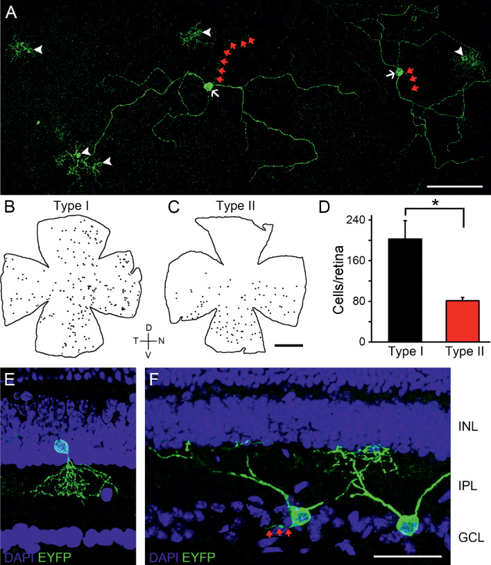
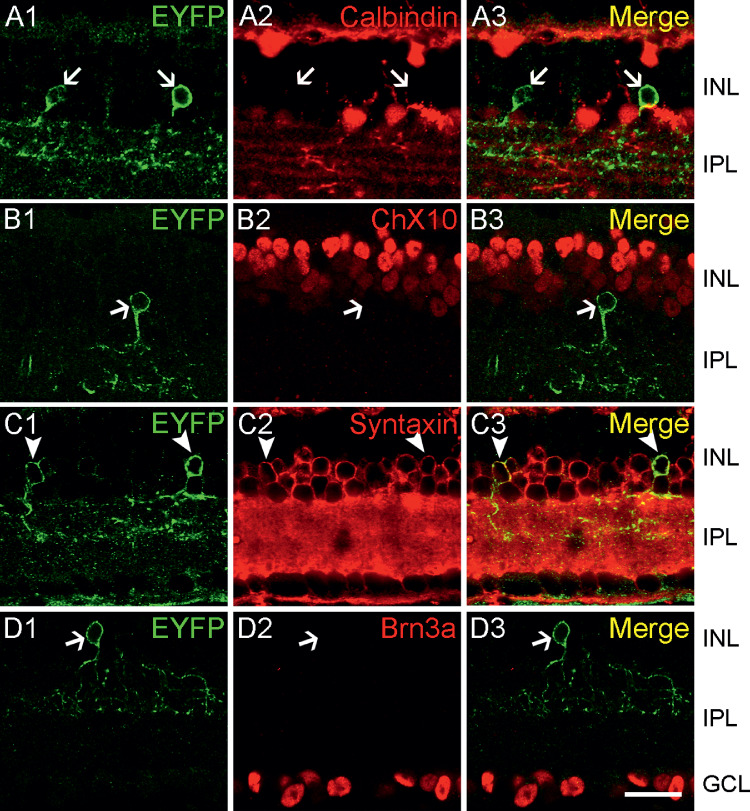
Two populations of EYFP-positive retinal cells were observed. The inner nuclear layer (INL)-located one (type I cell) distributed regularly throughout the entire retina, whereas the ganglion cell layer (GCL)-residing one (type II cell) was restricted ventrally. None of them was cholinergic, as evidenced by the complete absence of ChAT immunoreactivity. Type I cells were immunolabeled by the amacrine marker syntaxin. However, the vast majority of them were neither positive to GABA/GAD65, nor to GlyT1/glycine, suggesting that they were non-GABAergic non-glycinergic amacrine cells (nGnG ACs), which was confirmed by double-labeling with the nGnG AC marker PPP1R17. Type II cells were immunopositive to melanopsin, but not to Brn3a or Brn3b. They possessed dendrites stratifying in the outermost inner plexiform layer (IPL) and axons projecting to the suprachiasmatic nucleus (SCN) rather than the olivary pretectal nucleus (OPN), suggesting that they belonged to a Brn3b-negative subset of M1-type intrinsically photosensitive retinal ganglion cells (ipRGCs). Glutamatergic transmission-independent photocurrents were elicited in EYFP-positive cells, indicating the functional expression of ChR2.
The ChAT-ChR2-EYFP retina exhibits ectopic, but functional, transgene expression in nGnG ACs and SCN-innervating M1 ipRGCs, thus providing an ideal tool to achieve efficient labeling and optogenetic manipulation of these cells.
实验性地获取特定细胞亚型对于解析视网膜网络的复杂性至关重要。在这里,我们对 ChAT-Channelrhodopsin-2 (ChR2)-EYFP 小鼠中两种非典型视网膜神经元的异位转基因表达所导致的选择性标记进行了描述。
制备视网膜切片和平面培养物,并用针对 EYFP 和各种神经元标志物的抗体进行双重免疫组织化学染色。制备矢状/冠状脑切片以可视化中枢核团中的 EYFP 信号。进行全细胞记录以测试 ChR2 的功能。
观察到两种 EYFP 阳性视网膜细胞群体。位于内核层 (INL) 的一种(I 型细胞)在整个视网膜中均匀分布,而位于神经节细胞层 (GCL) 的一种(II 型细胞)则局限于腹侧。它们都不是胆碱能的,因为它们完全缺乏 ChAT 免疫反应性。I 型细胞被突触素免疫标记。然而,它们中的绝大多数既不表达 GABA/GAD65,也不表达 GlyT1/甘氨酸,表明它们是非 GABA 能非甘氨酸能无长突细胞 (nGnG ACs),这一点通过与 nGnG AC 标志物 PPP1R17 的双重标记得到了证实。II 型细胞对黑视素呈免疫阳性,但对 Brn3a 或 Brn3b 呈阴性。它们的树突分层于最外层的内丛状层 (IPL),轴突投射到视交叉上核 (SCN),而不是橄榄状视前核 (OPN),表明它们属于 Brn3b 阴性的 M1 型内在光敏性视网膜神经节细胞 (ipRGCs) 亚群。在 EYFP 阳性细胞中引发了谷氨酸能传递非依赖性光电流,表明 ChR2 的功能表达。
ChAT-ChR2-EYFP 视网膜在 nGnG AC 和 SCN 支配的 M1 ipRGC 中表现出异位但功能性的转基因表达,因此提供了一种理想的工具,可以有效地标记和光遗传学操纵这些细胞。