• 文献检索
  • 文档翻译
  • 深度研究
  • 学术资讯
  • Suppr Zotero 插件Zotero 插件
  • 邀请有礼
  • 套餐&价格
  • 历史记录
应用&插件
Suppr Zotero 插件Zotero 插件浏览器插件Mac 客户端Windows 客户端微信小程序
定价
高级版会员购买积分包购买API积分包
服务
文献检索文档翻译深度研究API 文档MCP 服务
关于我们
关于 Suppr公司介绍联系我们用户协议隐私条款
关注我们

Suppr 超能文献

核心技术专利:CN118964589B侵权必究
粤ICP备2023148730 号-1Suppr @ 2026

文献检索

告别复杂PubMed语法,用中文像聊天一样搜索,搜遍4000万医学文献。AI智能推荐,让科研检索更轻松。

立即免费搜索

文件翻译

保留排版,准确专业,支持PDF/Word/PPT等文件格式,支持 12+语言互译。

免费翻译文档

深度研究

AI帮你快速写综述,25分钟生成高质量综述,智能提取关键信息,辅助科研写作。

立即免费体验

连接蛋白-2 将 T 小管固定在成年心肌细胞的脂筏上,并募集功能性 L 型钙通道。

Junctophilin-2 tethers T-tubules and recruits functional L-type calcium channels to lipid rafts in adult cardiomyocytes.

机构信息

National Heart and Lung Institute, Imperial College London, Du Cane Road, London W12 0NN, UK.

Department of Cardiac Surgery, School of Medicine, University of Verona, Piazzale L.A. Scuro 10, 37134 Verona, Italy.

出版信息

Cardiovasc Res. 2021 Jan 1;117(1):149-161. doi: 10.1093/cvr/cvaa033.

DOI:10.1093/cvr/cvaa033
PMID:32053184
原文链接:https://pmc.ncbi.nlm.nih.gov/articles/PMC7797210/
Abstract

AIM

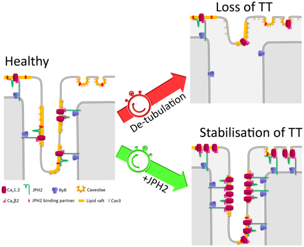
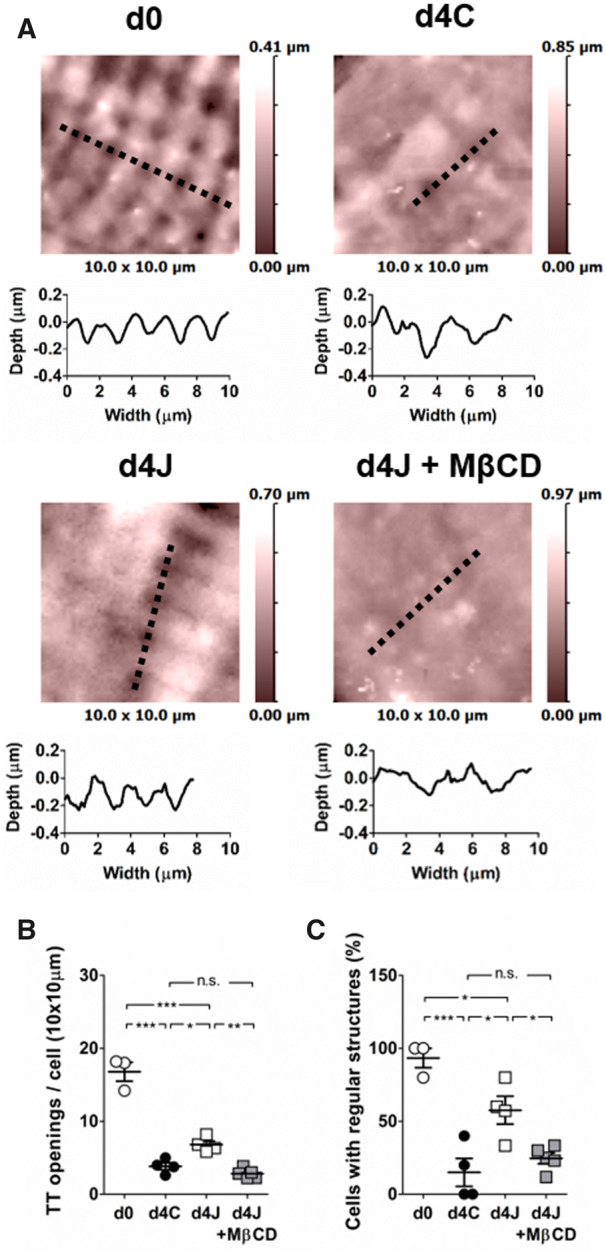
In cardiomyocytes, transverse tubules (T-tubules) associate with the sarcoplasmic reticulum (SR), forming junctional membrane complexes (JMCs) where L-type calcium channels (LTCCs) are juxtaposed to Ryanodine receptors (RyR). Junctophilin-2 (JPH2) supports the assembly of JMCs by tethering T-tubules to the SR membrane. T-tubule remodelling in cardiac diseases is associated with downregulation of JPH2 expression suggesting that JPH2 plays a crucial role in T-tubule stability. Furthermore, increasing evidence indicate that JPH2 might additionally act as a modulator of calcium signalling by directly regulating RyR and LTCCs. This study aimed at determining whether JPH2 overexpression restores normal T-tubule structure and LTCC function in cultured cardiomyocytes.

METHODS AND RESULTS

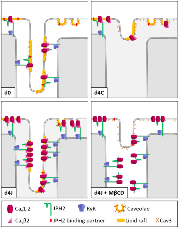
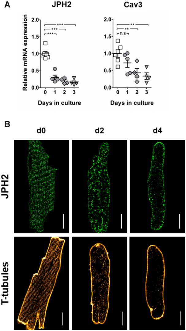
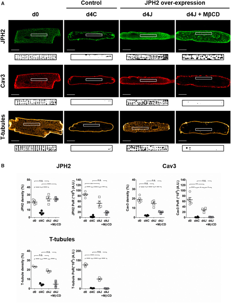
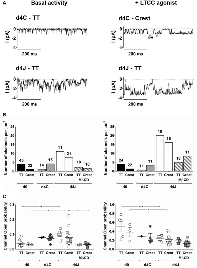
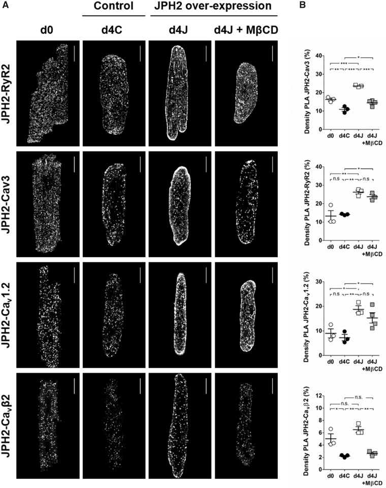
Rat ventricular myocytes kept in culture for 4 days showed extensive T-tubule remodelling with impaired JPH2 localization and relocation of the scaffolding protein Caveolin3 (Cav3) from the T-tubules to the outer membrane. Overexpression of JPH2 restored T-tubule structure and Cav3 relocation. Depletion of membrane cholesterol by chronic treatment with methyl-β-cyclodextrin (MβCD) countered the stabilizing effect of JPH2 overexpression on T-tubules and Cav3. Super-resolution scanning patch-clamp showed that JPH2 overexpression greatly increased the number of functional LTCCs at the plasma membrane. Treatment with MβCD reduced LTCC open probability and activity. Proximity ligation assays showed that MβCD did not affect JPH2 interaction with RyR and the pore-forming LTCC subunit Cav1.2, but strongly impaired JPH2 association with Cav3 and the accessory LTCC subunit Cavβ2.

CONCLUSIONS

JPH2 promotes T-tubule structural stability and recruits functional LTCCs to the membrane, most likely by directly binding to the channel. Cholesterol is involved in the binding of JPH2 to T-tubules as well as in the modulation of LTCC activity. We propose a model where cholesterol and Cav3 support the assembly of lipid rafts which provide an anchor for JPH2 to form JMCs and a platform for signalling complexes to regulate LTCC activity.

摘要

目的

在心肌细胞中,横管(T 管)与肌质网(SR)相连,形成连接膜复合物(JMC),其中 L 型钙通道(LTCC)与 Ryanodine 受体(RyR)并列。Junctophilin-2(JPH2)通过将 T 管固定在 SR 膜上,支持 JMC 的组装。心脏疾病中的 T 管重塑与 JPH2 表达下调有关,表明 JPH2 在 T 管稳定性中起关键作用。此外,越来越多的证据表明,JPH2 可能通过直接调节 RyR 和 LTCC 来充当钙信号转导的调节剂。本研究旨在确定 JPH2 的过表达是否能恢复培养的心肌细胞中正常的 T 管结构和 LTCC 功能。

方法和结果

在培养 4 天的大鼠心室肌细胞中,T 管广泛重塑,JPH2 定位受损,支架蛋白 Caveolin3(Cav3)从 T 管转移到外膜。JPH2 的过表达恢复了 T 管结构和 Cav3 的重定位。用甲基-β-环糊精(MβCD)慢性处理以耗竭膜胆固醇,抵消了 JPH2 过表达对 T 管和 Cav3 的稳定作用。超分辨率扫描贴片钳显示,JPH2 的过表达大大增加了质膜上功能性 LTCC 的数量。MβCD 的处理降低了 LTCC 的开放概率和活性。接近连接测定表明,MβCD 不影响 JPH2 与 RyR 和形成孔的 LTCC 亚基 Cav1.2 的相互作用,但强烈抑制了 JPH2 与 Cav3 和辅助 LTCC 亚基 Cavβ2 的结合。

结论

JPH2 促进 T 管结构稳定性,并将功能性 LTCC 募集到膜上,这很可能是通过直接与通道结合。胆固醇参与 JPH2 与 T 管的结合以及 LTCC 活性的调节。我们提出了一个模型,其中胆固醇和 Cav3 支持脂筏的组装,脂筏为 JPH2 形成 JMC 提供了一个锚点,并为信号复合物调节 LTCC 活性提供了一个平台。

https://cdn.ncbi.nlm.nih.gov/pmc/blobs/4dfb/7797210/9446fbeff713/cvaa033f7.jpg
https://cdn.ncbi.nlm.nih.gov/pmc/blobs/4dfb/7797210/f67a265a172f/cvaa033f8.jpg
https://cdn.ncbi.nlm.nih.gov/pmc/blobs/4dfb/7797210/6e98947ccc47/cvaa033f1.jpg
https://cdn.ncbi.nlm.nih.gov/pmc/blobs/4dfb/7797210/cf521ca176b3/cvaa033f2.jpg
https://cdn.ncbi.nlm.nih.gov/pmc/blobs/4dfb/7797210/af74081df405/cvaa033f3.jpg
https://cdn.ncbi.nlm.nih.gov/pmc/blobs/4dfb/7797210/5c3d1c5198a0/cvaa033f4.jpg
https://cdn.ncbi.nlm.nih.gov/pmc/blobs/4dfb/7797210/668b849b97f0/cvaa033f5.jpg
https://cdn.ncbi.nlm.nih.gov/pmc/blobs/4dfb/7797210/f13e4a4e383b/cvaa033f6.jpg
https://cdn.ncbi.nlm.nih.gov/pmc/blobs/4dfb/7797210/9446fbeff713/cvaa033f7.jpg

相似文献

1
Junctophilin-2 tethers T-tubules and recruits functional L-type calcium channels to lipid rafts in adult cardiomyocytes.连接蛋白-2 将 T 小管固定在成年心肌细胞的脂筏上,并募集功能性 L 型钙通道。
Cardiovasc Res. 2021 Jan 1;117(1):149-161. doi: 10.1093/cvr/cvaa033.
2
Interaction of the Joining Region in Junctophilin-2 With the L-Type Ca Channel Is Pivotal for Cardiac Dyad Assembly and Intracellular Ca Dynamics.衔接蛋白 Junctophilin-2 的连接区与 L 型钙通道的相互作用对于心肌二联体的组装和细胞内钙动力学至关重要。
Circ Res. 2021 Jan 8;128(1):92-114. doi: 10.1161/CIRCRESAHA.119.315715. Epub 2020 Oct 23.
3
Direct Evidence for Microdomain-Specific Localization and Remodeling of Functional L-Type Calcium Channels in Rat and Human Atrial Myocytes.大鼠和人类心房肌细胞中功能性L型钙通道微区特异性定位和重塑的直接证据
Circulation. 2015 Dec 22;132(25):2372-84. doi: 10.1161/CIRCULATIONAHA.115.018131. Epub 2015 Oct 8.
4
Junctophilin-2 gene therapy rescues heart failure by normalizing RyR2-mediated Ca release.连接蛋白-2基因疗法通过使兰尼碱受体2介导的钙释放正常化来挽救心力衰竭。
Int J Cardiol. 2016 Dec 15;225:371-380. doi: 10.1016/j.ijcard.2016.10.021. Epub 2016 Oct 8.
5
Caveolin 3-dependent loss of t-tubular I during hypertrophy and heart failure in mice.小鼠肥大和心力衰竭过程中,小窝蛋白3依赖性横管肌浆网连接蛋白I的缺失
Exp Physiol. 2018 May 1;103(5):652-665. doi: 10.1113/EP086731. Epub 2018 Apr 14.
6
Functional LTCC-βAR Complex Needs Caveolin-3 and Is Disrupted in Heart Failure.功能性 LTCC-βAR 复合物需要 caveolin-3 并在心力衰竭中被破坏。
Circ Res. 2023 Jul 7;133(2):120-137. doi: 10.1161/CIRCRESAHA.123.322508. Epub 2023 Jun 14.
7
Nanoscale organization of cardiac calcium channels is dependent on thyroid hormone status.心脏钙通道的纳米级组织依赖于甲状腺激素状态。
Am J Physiol Heart Circ Physiol. 2024 Nov 1;327(5):H1309-H1326. doi: 10.1152/ajpheart.00272.2024. Epub 2024 Oct 4.
8
Triiodothyronine maintains cardiac transverse-tubule structure and function.三碘甲状腺原氨酸维持心脏横管结构和功能。
J Mol Cell Cardiol. 2021 Nov;160:1-14. doi: 10.1016/j.yjmcc.2021.06.010. Epub 2021 Jun 24.
9
T-tubule remodelling disturbs localized β2-adrenergic signalling in rat ventricular myocytes during the progression of heart failure.在心力衰竭进展过程中,横管重塑会干扰大鼠心室肌细胞中局部的β2-肾上腺素能信号传导。
Cardiovasc Res. 2017 Jun 1;113(7):770-782. doi: 10.1093/cvr/cvx074.
10
BIN1 regulates dynamic t-tubule membrane.BIN1调节动态横管膜。
Biochim Biophys Acta. 2016 Jul;1863(7 Pt B):1839-47. doi: 10.1016/j.bbamcr.2015.11.004. Epub 2015 Nov 11.

引用本文的文献

1
Role of Dyadic Proteins in Proper Heart Function and Disease.二元蛋白在心脏正常功能和疾病中的作用。
Int J Mol Sci. 2025 Aug 2;26(15):7478. doi: 10.3390/ijms26157478.
2
Ca microdomain-based excitation-transcription coupling in cardiac myocytes and vascular smooth muscle cells.心肌细胞和血管平滑肌细胞中基于钙微区的兴奋-转录偶联
Inflamm Regen. 2025 Jun 23;45(1):19. doi: 10.1186/s41232-025-00384-3.
3
Functional analysis of JPH2-knockout cardiomyocytes identifies ECCD as a novel indicator in a human cardiac modelJPH2.JPH2基因敲除心肌细胞的功能分析确定ECCD是人类心脏模型JPH2中的一种新指标。

本文引用的文献

1
Cardiomyocyte substructure reverts to an immature phenotype during heart failure.在心力衰竭过程中心肌细胞亚结构恢复为未成熟表型。
J Physiol. 2019 Apr;597(7):1833-1853. doi: 10.1113/JP277273. Epub 2019 Feb 27.
2
Cardiac CaV1.2 channels require β subunits for β-adrenergic-mediated modulation but not trafficking.心肌 CaV1.2 通道需要β亚基来进行β肾上腺素能介导的调节,但不需要运输。
J Clin Invest. 2019 Feb 1;129(2):647-658. doi: 10.1172/JCI123878. Epub 2019 Jan 7.
3
E-C coupling structural protein junctophilin-2 encodes a stress-adaptive transcription regulator.
Stem Cell Res Ther. 2025 May 9;16(1):234. doi: 10.1186/s13287-025-04323-4.
4
A 5-lncRNA signature predicts clinical prognosis and demonstrates a different mRNA expression in adult soft tissue sarcoma.一种5-长链非编码RNA特征可预测临床预后,并在成人软组织肉瘤中表现出不同的mRNA表达。
Transl Cancer Res. 2025 Jan 31;14(1):179-196. doi: 10.21037/tcr-24-203. Epub 2025 Jan 23.
5
Constitutive deletion of the obscurin-Ig58/59 domains induces atrial remodeling and Ca2+-based arrhythmogenesis.obscurin-Ig58/59结构域的组成性缺失会诱导心房重塑和基于Ca2+的心律失常发生。
JCI Insight. 2025 Jan 7;10(4):e184202. doi: 10.1172/jci.insight.184202.
6
Channels, Transporters, and Receptors at Membrane Contact Sites.膜接触位点处的通道、转运体和受体
Contact (Thousand Oaks). 2024 Dec 26;7:25152564241305593. doi: 10.1177/25152564241305593. eCollection 2024 Jan-Dec.
7
Preventing Site-Specific Calpain Proteolysis of Junctophilin-2 Protects Against Stress-Induced Excitation-Contraction Uncoupling and Heart Failure Development.防止连接蛋白2的位点特异性钙蛋白酶解可预防应激诱导的兴奋-收缩偶联障碍和心力衰竭发展。
Circulation. 2025 Jan 14;151(2):171-187. doi: 10.1161/CIRCULATIONAHA.124.069329. Epub 2024 Sep 18.
8
FOXA3 regulates cholesterol metabolism to compensate for low uptake during the progression of lung adenocarcinoma.FOXA3 通过调节胆固醇代谢来补偿肺腺癌进展过程中的摄取减少。
PLoS Biol. 2024 May 28;22(5):e3002621. doi: 10.1371/journal.pbio.3002621. eCollection 2024 May.
9
Caveolar Compartmentalization of Pacemaker Signaling is Required for Stable Rhythmicity of Sinus Nodal Cells and is Disrupted in Heart Failure.窦房结细胞稳定节律性需要起搏器信号的小窝区室化,且心力衰竭时该过程会受到破坏。
bioRxiv. 2024 Aug 27:2024.04.14.589457. doi: 10.1101/2024.04.14.589457.
10
Altered myocardial lipid regulation in junctophilin-2-associated familial cardiomyopathies.肌联蛋白-2 相关家族性心肌病中心肌脂质调节的改变。
Life Sci Alliance. 2024 Mar 4;7(5). doi: 10.26508/lsa.202302330. Print 2024 May.
E-C 偶联结构蛋白连接蛋白-2 编码一种应激适应性转录调节因子。
Science. 2018 Dec 21;362(6421). doi: 10.1126/science.aan3303. Epub 2018 Nov 8.
4
Physical interaction of junctophilin and the Ca1.1 C terminus is crucial for skeletal muscle contraction.连接蛋白和 Ca1.1 C 端的物理相互作用对于骨骼肌收缩至关重要。
Proc Natl Acad Sci U S A. 2018 Apr 24;115(17):4507-4512. doi: 10.1073/pnas.1716649115. Epub 2018 Apr 9.
5
True Molecular Scale Visualization of Variable Clustering Properties of Ryanodine Receptors.真分子水平可视化兰尼碱受体的变构簇性质。
Cell Rep. 2018 Jan 9;22(2):557-567. doi: 10.1016/j.celrep.2017.12.045.
6
T-tubule remodelling disturbs localized β2-adrenergic signalling in rat ventricular myocytes during the progression of heart failure.在心力衰竭进展过程中,横管重塑会干扰大鼠心室肌细胞中局部的β2-肾上腺素能信号传导。
Cardiovasc Res. 2017 Jun 1;113(7):770-782. doi: 10.1093/cvr/cvx074.
7
Analysis of Cardiac Myocyte Maturation Using CASAAV, a Platform for Rapid Dissection of Cardiac Myocyte Gene Function In Vivo.使用CASAAV分析心肌细胞成熟,CASAAV是一种用于在体内快速剖析心肌细胞基因功能的平台。
Circ Res. 2017 Jun 9;120(12):1874-1888. doi: 10.1161/CIRCRESAHA.116.310283. Epub 2017 Mar 29.
8
Junctophilin-2 in the nanoscale organisation and functional signalling of ryanodine receptor clusters in cardiomyocytes.连接蛋白2在心肌细胞中兰尼碱受体簇的纳米级组织和功能信号传导中的作用
J Cell Sci. 2016 Dec 1;129(23):4388-4398. doi: 10.1242/jcs.196873. Epub 2016 Oct 21.
9
Junctophilin-2 gene therapy rescues heart failure by normalizing RyR2-mediated Ca release.连接蛋白-2基因疗法通过使兰尼碱受体2介导的钙释放正常化来挽救心力衰竭。
Int J Cardiol. 2016 Dec 15;225:371-380. doi: 10.1016/j.ijcard.2016.10.021. Epub 2016 Oct 8.
10
Microdomain-Specific Modulation of L-Type Calcium Channels Leads to Triggered Ventricular Arrhythmia in Heart Failure.L型钙通道的微区特异性调节导致心力衰竭时的触发型室性心律失常。
Circ Res. 2016 Sep 30;119(8):944-55. doi: 10.1161/CIRCRESAHA.116.308698. Epub 2016 Aug 29.