• 文献检索
  • 文档翻译
  • 深度研究
  • 学术资讯
  • Suppr Zotero 插件Zotero 插件
  • 邀请有礼
  • 套餐&价格
  • 历史记录
应用&插件
Suppr Zotero 插件Zotero 插件浏览器插件Mac 客户端Windows 客户端微信小程序
定价
高级版会员购买积分包购买API积分包
服务
文献检索文档翻译深度研究API 文档MCP 服务
关于我们
关于 Suppr公司介绍联系我们用户协议隐私条款
关注我们

Suppr 超能文献

核心技术专利:CN118964589B侵权必究
粤ICP备2023148730 号-1Suppr @ 2026

文献检索

告别复杂PubMed语法,用中文像聊天一样搜索,搜遍4000万医学文献。AI智能推荐,让科研检索更轻松。

立即免费搜索

文件翻译

保留排版,准确专业,支持PDF/Word/PPT等文件格式,支持 12+语言互译。

免费翻译文档

深度研究

AI帮你快速写综述,25分钟生成高质量综述,智能提取关键信息,辅助科研写作。

立即免费体验

斑状暗点见于近视性黄斑变性的脉络膜视网膜斑状萎缩。

Patchy Scotoma Observed in Chorioretinal Patchy Atrophy of Myopic Macular Degeneration.

机构信息

,.

出版信息

Invest Ophthalmol Vis Sci. 2020 Feb 7;61(2):15. doi: 10.1167/iovs.61.2.15.

DOI:10.1167/iovs.61.2.15
PMID:32053726
原文链接:https://pmc.ncbi.nlm.nih.gov/articles/PMC7326503/
Abstract

PURPOSE

To investigate the retinal sensitivity of highly myopic eyes with chorioretinal patchy atrophy (PA) using microperimetry.

METHODS

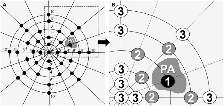
Fifty-two eyes of 32 patients with high myopia were prospectively included. Twenty-two eyes of 16 patients had PA lesions; eyes without PA were analyzed as controls. Testing points on microperimetry in eyes with PA were designated as 3 zones: zone 1 as the PA lesion including its borders; zone 2 including testing points adjoining PA; zone 3 including all other testing points.

RESULTS

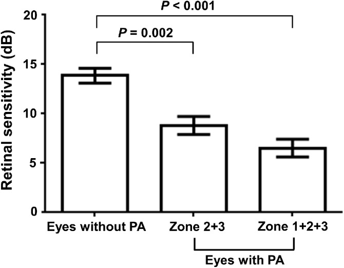
In the PA group, the mean retinal sensitivity in zone 1 was 2.1 ± 2.8 dB, zone 2 = 8.3 ± 4.3 dB, and zone 3 = 9.4 ± 4.1 dB. Sensitivity in zone 1 was significantly reduced than zones 2 and 3 (P < 0.001). The mean retinal sensitivity in the PA group was lower than controls (6.5 ± 4.3 vs 13.9 ± 4.1 dB, P < 0.001), and combined zone 2 and 3 in the PA group also presented lower retinal sensitivity (8.8 ± 4.0 dB).

CONCLUSIONS

Eyes with PA generate patchy scotoma in PA lesions and reduced retinal sensitivity in regions beyond atrophic lesion on microperimetry. The presence of PA may be an indicator to reflect both significantly anatomical and functional alterations on myopic macular degeneration.

摘要

目的

使用微视野计研究伴有脉络膜视网膜斑状萎缩(PA)的高度近视眼的视网膜敏感性。

方法

前瞻性纳入 32 名患者的 52 只高度近视眼。22 只眼有 PA 病变;无 PA 的眼作为对照进行分析。PA 眼的微视野计检测点被指定为 3 个区:区 1 为包括边界的 PA 病变;区 2 包括毗邻 PA 的检测点;区 3 包括所有其他检测点。

结果

在 PA 组中,区 1 的平均视网膜敏感性为 2.1 ± 2.8 dB,区 2 = 8.3 ± 4.3 dB,区 3 = 9.4 ± 4.1 dB。区 1 的敏感性明显低于区 2 和区 3(P < 0.001)。PA 组的平均视网膜敏感性低于对照组(6.5 ± 4.3 与 13.9 ± 4.1 dB,P < 0.001),PA 组的区 2 和区 3 联合敏感性也较低(8.8 ± 4.0 dB)。

结论

PA 眼在 PA 病变中产生斑状暗点,在微视野计上萎缩病变之外的区域视网膜敏感性降低。PA 的存在可能是反映近视性黄斑变性的显著解剖和功能改变的指标。

https://cdn.ncbi.nlm.nih.gov/pmc/blobs/0238/7326503/2f3950279c75/iovs-61-2-15-f004.jpg
https://cdn.ncbi.nlm.nih.gov/pmc/blobs/0238/7326503/6bf53d56908a/iovs-61-2-15-f001.jpg
https://cdn.ncbi.nlm.nih.gov/pmc/blobs/0238/7326503/4191365e21e7/iovs-61-2-15-f002.jpg
https://cdn.ncbi.nlm.nih.gov/pmc/blobs/0238/7326503/30005e53c2a8/iovs-61-2-15-f003.jpg
https://cdn.ncbi.nlm.nih.gov/pmc/blobs/0238/7326503/2f3950279c75/iovs-61-2-15-f004.jpg

相似文献

1
Patchy Scotoma Observed in Chorioretinal Patchy Atrophy of Myopic Macular Degeneration.斑状暗点见于近视性黄斑变性的脉络膜视网膜斑状萎缩。
Invest Ophthalmol Vis Sci. 2020 Feb 7;61(2):15. doi: 10.1167/iovs.61.2.15.
2
Macular Bruch Membrane Holes in Highly Myopic Patchy Chorioretinal Atrophy.高度近视性斑片状脉络膜视网膜萎缩中的黄斑布鲁赫膜孔
Am J Ophthalmol. 2016 Jun;166:22-28. doi: 10.1016/j.ajo.2016.03.019. Epub 2016 Mar 24.
3
Association of Retinal Sensitivity With Optical Coherence Tomography Microstructure in Highly Myopic Patients.高度近视患者视网膜敏感度与光学相干断层扫描微观结构的相关性。
Invest Ophthalmol Vis Sci. 2022 Oct 3;63(11):13. doi: 10.1167/iovs.63.11.13.
4
Evolution of Macular Bruch Membrane Defects of Patchy Chorioretinal Atrophy in Pathologic Myopia Based on a Recent Classification System.基于最近的分类系统探讨病理性近视中斑状脉络膜视网膜萎缩的脉络膜视网膜玻璃膜疣的演变。
Ophthalmologica. 2021;244(4):309-314. doi: 10.1159/000516526. Epub 2021 May 3.
5
ABRUPTLY EMERGING VESSELS IN EYES WITH MYOPIC PATCHY CHORIORETINAL ATROPHY.伴有近视性斑状脉络膜视网膜萎缩的突然出现的眼底血管。
Retina. 2020 Jul;40(7):1215-1223. doi: 10.1097/IAE.0000000000002630.
6
Retinal Sensitivity at the Junctional Zone of Eyes With Geographic Atrophy Due to Age-Related Macular Degeneration.年龄相关性黄斑变性所致地图样萎缩眼中连接区的视网膜敏感性
Am J Ophthalmol. 2016 Aug;168:122-128. doi: 10.1016/j.ajo.2016.05.007. Epub 2016 May 14.
7
Macular Bruch Membrane Holes in Choroidal Neovascularization-Related Myopic Macular Atrophy by Swept-Source Optical Coherence Tomography.扫频光学相干断层扫描检测脉络膜新生血管相关近视性黄斑萎缩中的黄斑 Bruch 膜孔
Am J Ophthalmol. 2016 Feb;162:133-139.e1. doi: 10.1016/j.ajo.2015.11.014. Epub 2015 Nov 14.
8
Progression and associated factors of lacquer cracks/patchy atrophies in high myopia: the Beijing Eye Study 2001-2011.高度近视的漆裂纹/斑状萎缩进展及其相关因素:2001-2011 年北京眼研究。
Graefes Arch Clin Exp Ophthalmol. 2022 Oct;260(10):3221-3229. doi: 10.1007/s00417-022-05705-7. Epub 2022 May 24.
9
Fundus autofluorescence and microperimetry in progressing geographic atrophy secondary to age-related macular degeneration.年龄相关性黄斑变性继发进行性地图状萎缩的眼底自发荧光和微视野检查。
Br J Ophthalmol. 2013 May;97(5):622-6. doi: 10.1136/bjophthalmol-2012-302633. Epub 2013 Feb 14.
10
Central retinal sensitivity after intravitreal injection of bevacizumab for myopic choroidal neovascularization.玻璃体内注射贝伐单抗治疗近视性脉络膜新生血管后的中央视网膜敏感度
Am J Ophthalmol. 2009 May;147(5):816-24, 824.e1. doi: 10.1016/j.ajo.2008.11.020. Epub 2009 Feb 10.

引用本文的文献

1
Association of Retinal Sensitivity With Optical Coherence Tomography Microstructure in Highly Myopic Patients.高度近视患者视网膜敏感度与光学相干断层扫描微观结构的相关性。
Invest Ophthalmol Vis Sci. 2022 Oct 3;63(11):13. doi: 10.1167/iovs.63.11.13.
2
IMI Pathologic Myopia.病理性近视
Invest Ophthalmol Vis Sci. 2021 Apr 28;62(5):5. doi: 10.1167/iovs.62.5.5.

本文引用的文献

1
Changes in Choroidal Thickness and Choroidal Blood Perfusion in Guinea Pig Myopia.豚鼠近视模型脉络膜厚度和脉络膜血流的变化。
Invest Ophthalmol Vis Sci. 2019 Jul 1;60(8):3074-3083. doi: 10.1167/iovs.18-26397.
2
OCT-Based Diagnostic Criteria for Different Stages of Myopic Maculopathy.基于 OCT 的不同阶段近视性黄斑病变的诊断标准。
Ophthalmology. 2019 Jul;126(7):1018-1032. doi: 10.1016/j.ophtha.2019.01.012. Epub 2019 Jan 29.
3
Progression of Myopic Maculopathy during 18-Year Follow-up.近视性黄斑病变 18 年随访进展。
Ophthalmology. 2018 Jun;125(6):863-877. doi: 10.1016/j.ophtha.2017.12.005. Epub 2018 Jan 19.
4
EVALUATION OF PATCHY ATROPHY SECONDARY TO HIGH MYOPIA BY SEMIAUTOMATED SOFTWARE FOR FUNDUS AUTOFLUORESCENCE ANALYSIS.应用眼底自发荧光分析半自动软件评估高度近视的斑状萎缩。
Retina. 2018 Jul;38(7):1301-1306. doi: 10.1097/IAE.0000000000001733.
5
The Use of Microperimetry to Detect Functional Progression in Non-Neovascular Age-Related Macular Degeneration: A Systematic Review.使用微视野检查法检测非新生血管性年龄相关性黄斑变性的功能进展:一项系统评价
Asia Pac J Ophthalmol (Phila). 2017 Jan-Feb;6(1):70-79. doi: 10.22608/APO.201643.
6
Macular Bruch Membrane Holes in Highly Myopic Patchy Chorioretinal Atrophy.高度近视性斑片状脉络膜视网膜萎缩中的黄斑布鲁赫膜孔
Am J Ophthalmol. 2016 Jun;166:22-28. doi: 10.1016/j.ajo.2016.03.019. Epub 2016 Mar 24.
7
International photographic classification and grading system for myopic maculopathy.近视性黄斑病变国际摄影分类及分级系统
Am J Ophthalmol. 2015 May;159(5):877-83.e7. doi: 10.1016/j.ajo.2015.01.022. Epub 2015 Jan 26.
8
Longitudinal changes in microperimetry and low luminance visual acuity in age-related macular degeneration.年龄相关性黄斑变性的微视野和低亮度视力的纵向变化。
JAMA Ophthalmol. 2015 Apr;133(4):442-8. doi: 10.1001/jamaophthalmol.2014.5963.
9
Retinal sensitivity and choroidal thickness in high myopia.高度近视中的视网膜敏感性和脉络膜厚度
Retina. 2015 Mar;35(3):398-406. doi: 10.1097/IAE.0000000000000367.
10
Microperimetry and clinical practice: an evidence-based review.微视野与临床实践:基于证据的综述。
Can J Ophthalmol. 2013 Oct;48(5):350-7. doi: 10.1016/j.jcjo.2012.03.004. Epub 2012 Oct 23.