Knight Cardiovascular Institute, Oregon Health & Science University, Portland, OR, 97239, USA.
Cardiovascular Division, Knight Cardiovascular Institute, The Oregon National Primate Research Center, Oregon Health & Science University, 3181 SW Sam Jackson Park Rd, Portland, OR, 97239, USA.
J Echocardiogr. 2020 Jun;18(2):86-93. doi: 10.1007/s12574-020-00463-z. Epub 2020 Feb 13.
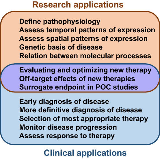
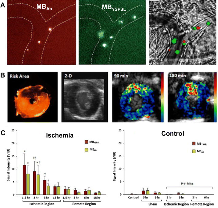
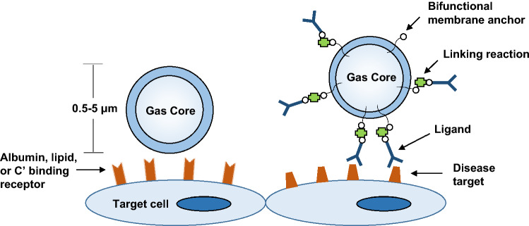
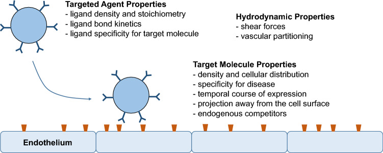
Similar to what has already occurred in cancer medicine, the management of cardiovascular conditions will likely be improved by non-invasive molecular imaging technologies that can provide earlier or more accurate diagnosis. These techniques are already having a positive impact in pre-clinical research by providing insight into pathophysiology or efficacy of new therapies. Contrast enhanced ultrasound (CEU) molecular imaging is a technique that relies on the ultrasound detection of targeted microbubble contrast agents to examine molecular or cellular events that occur at the blood pool-endothelial interface. CEU molecular imaging techniques have been developed that are able to provide unique information on atherosclerosis, ischemia reperfusion injury, angiogenesis, vascular inflammation, and thrombus formation. Accordingly, CEU has the potential to be used in a wide variety of circumstances to detect disease early or at the bedside, and to guide appropriate therapy based on vascular phenotype. This review will describe the physical basis for CEU molecular imaging, and the specific disease processes for the pre-clinical translational research experience.
类似于癌症医学中已经发生的情况,通过提供更早或更准确的诊断的非侵入性分子成像技术,心血管疾病的治疗可能会得到改善。这些技术已经通过提供对新疗法的病理生理学或疗效的深入了解,对临床前研究产生了积极影响。对比增强超声(CEU)分子成像技术是一种依赖于靶向微泡造影剂的超声检测来检查发生在血液池-内皮界面的分子或细胞事件的技术。已经开发出能够提供关于动脉粥样硬化、缺血再灌注损伤、血管生成、血管炎症和血栓形成的独特信息的 CEU 分子成像技术。因此,CEU 有可能在广泛的情况下用于早期检测疾病或在床边检测疾病,并根据血管表型指导适当的治疗。本综述将描述 CEU 分子成像的物理基础,以及临床前转化研究经验中的具体疾病过程。