Albalooshi Batool, Al Sharhan Mouza, Bagheri Fariborz, Miyanath Shabna, Ray Bhavna, Muhasin Muhammed, Zakavi Seyed Rasoul
Dubai Nuclear Medicine and Molecular imaging Center-Dubai Health Authority, Dubai, UAE.
Department of Genetics and Pathology-Dubai Health Authority, Dubai, UAE.
Asia Ocean J Nucl Med Biol. 2020 Winter;8(1):1-7. doi: 10.22038/aojnmb.2019.43943.1293.
Tc-PSMA SPECT/CT is a cost effective alternative for Ga-PSMA PET/CT. The aim of this study was to directly compare these two techniques in patients with prostate cancer.
28 man with prostate cancer were studied using Tc-PSMA SPECT/CT and Ga-PSMA PET/CT in a short time period (<60 days). No intervention was done between the studies. Whole body PET/CT was done 60 minutes after IV injection of 2 MBq/Kg of Ga-PSMA. Tc-PSMA kit (PSMA I+S) was used for SPECT/CT and whole body imaging was performed 4 hours after IV injection of 740 MBq of Tc-PSMA. Images were interpreted independently and the results of each imaging were recorded.
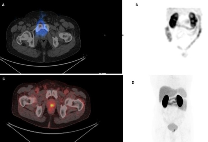
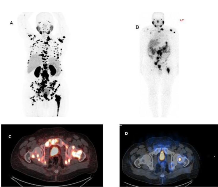
The mean age of the patients was 64.7±9.6 years old and the mean time difference between two sets of images was 16.6±13.5 days. Abnormal uptake was seen in 25 (89.2%) patients by Ga-PSMA PET/CT and 20 (71.4%) patients with Tc-PSMA SPECT/CT. No patients with positive Tc-PSMA SPECT/CT had negative Ga-PSMA PET/CT. The mean number of detected lesions was 26.07±27.5 by Ga-PSMA PET/CT and 10.52±10.99 by Tc-PSMA SPECT/CT (P<0.001). Detection of lymph nodes and bone metastases were not significantly different between two sets of imaging (P>0.05), however Ga-PSMA PET/CT were more successful in detection of prostate bed lesions compared to Tc-PSMA scan. Interestingly, no patient with PSA level of >2.1 ng/ml had discordant result between two sets of images.
Tc-PSMA SPECT/CT is as accurate as Ga-PSMA PET/CT in M staging, however Ga-PSMA PET/CT detected more lesions compared to Tc-PSMA SPECT/CT. Detection rate was not significantly different between two techniques in patients with PSA levels>2.1 ng/ml.
锝-PSMA SPECT/CT是镓-PSMA PET/CT一种性价比高的替代方法。本研究的目的是在前列腺癌患者中直接比较这两种技术。
在短时间内(<60天)对28名前列腺癌男性患者使用锝-PSMA SPECT/CT和镓-PSMA PET/CT进行研究。两次检查之间未进行干预。静脉注射2MBq/Kg的镓-PSMA后60分钟进行全身PET/CT检查。锝-PSMA试剂盒(PSMA I+S)用于SPECT/CT检查,静脉注射740MBq的锝-PSMA后4小时进行全身成像。图像由独立人员解读,并记录每次成像的结果。
患者的平均年龄为64.7±9.6岁,两组图像之间的平均时间间隔为16.6±13.5天。镓-PSMA PET/CT检查发现25例(89.2%)患者有异常摄取,锝-PSMA SPECT/CT检查发现20例(71.4%)患者有异常摄取。锝-PSMA SPECT/CT检查阳性的患者中没有镓-PSMA PET/CT检查阴性的。镓-PSMA PET/CT检查发现的病变平均数量为26.07±27.5个,锝-PSMA SPECT/CT检查发现的病变平均数量为10.52±10.99个(P<0.001)。两组成像在检测淋巴结和骨转移方面无显著差异(P>0.05),然而与锝-PSMA扫描相比,镓-PSMA PET/CT在检测前列腺床病变方面更成功。有趣的是,前列腺特异性抗原(PSA)水平>2.1ng/ml的患者中,两组图像之间没有不一致的结果。
在M分期方面,锝-PSMA SPECT/CT与镓-PSMA PET/CT一样准确,然而与锝-PSMA SPECT/CT相比,镓-PSMA PET/CT检测到的病变更多。在PSA水平>2.1ng/ml的患者中,两种技术的检测率无显著差异。