• 文献检索
  • 文档翻译
  • 深度研究
  • 学术资讯
  • Suppr Zotero 插件Zotero 插件
  • 邀请有礼
  • 套餐&价格
  • 历史记录
应用&插件
Suppr Zotero 插件Zotero 插件浏览器插件Mac 客户端Windows 客户端微信小程序
定价
高级版会员购买积分包购买API积分包
服务
文献检索文档翻译深度研究API 文档MCP 服务
关于我们
关于 Suppr公司介绍联系我们用户协议隐私条款
关注我们

Suppr 超能文献

核心技术专利:CN118964589B侵权必究
粤ICP备2023148730 号-1Suppr @ 2026

文献检索

告别复杂PubMed语法,用中文像聊天一样搜索,搜遍4000万医学文献。AI智能推荐,让科研检索更轻松。

立即免费搜索

文件翻译

保留排版,准确专业,支持PDF/Word/PPT等文件格式,支持 12+语言互译。

免费翻译文档

深度研究

AI帮你快速写综述,25分钟生成高质量综述,智能提取关键信息,辅助科研写作。

立即免费体验

前列腺癌患者骨转移的评估——锝骨闪烁显像与[镓]镓-PSMA PET/CT的比较

Assessment of Bone Metastases in Patients with Prostate Cancer-A Comparison between Tc-Bone-Scintigraphy and [Ga]Ga-PSMA PET/CT.

作者信息

Thomas Lena, Balmus Caroline, Ahmadzadehfar Hojjat, Essler Markus, Strunk Holger, Bundschuh Ralph A

机构信息

Klinik und Poliklinik für Nuklearmedizin, Universitätsklinikum Bonn, Bonn D-53127, Germany.

Radiologische Klinik, Universitätsklinikum Bonn, Bonn D-53012, Germany.

出版信息

Pharmaceuticals (Basel). 2017 Jul 31;10(3):68. doi: 10.3390/ph10030068.

DOI:10.3390/ph10030068
PMID:28758969
原文链接:https://pmc.ncbi.nlm.nih.gov/articles/PMC5620612/
Abstract

PURPOSE

Bone scintigraphy is the standard of reference in bone metastases in prostate cancer patients. However, new radiotracers employed in prostate-specific membrane antigen (PSMA)-ligands has led to the growing importance of PET/CT as diagnostic tool. The aim of our study was to investigate the difference between bone scan and PSMA-PET/CT for the detection of bone metastases in prostate cancer.

METHODS

Thirty patients with bone metastases originating from prostate cancer were examined by Tc-MDP bone scan and Ga-PSMA-PET/CT within an average of 21 days. Bone scans were analyzed visually according to the number of lesions and using the software package ExiniBONE by Exini Diagnostics. PET/CT data was analyzed visually. Numbers of detected lesions were compared for the different methods for the whole patient and for different regions. In addition, results were compared to serum prostate-specific antigen (PSA), alkaline phosphatase (ALP), bone alkaline phosphatase (bALP), pro gastrin releasing peptide (pGRP) and eastern cooperative oncology group (ECOG) performance status.

RESULTS

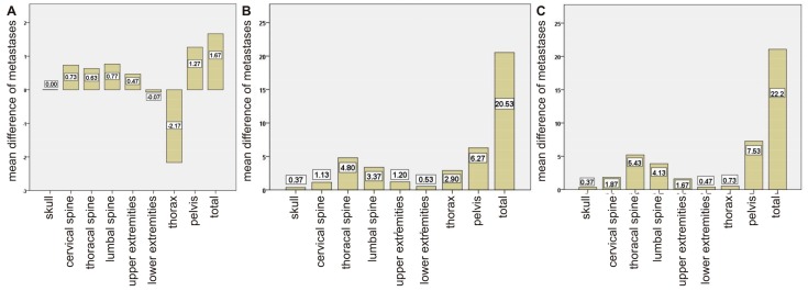
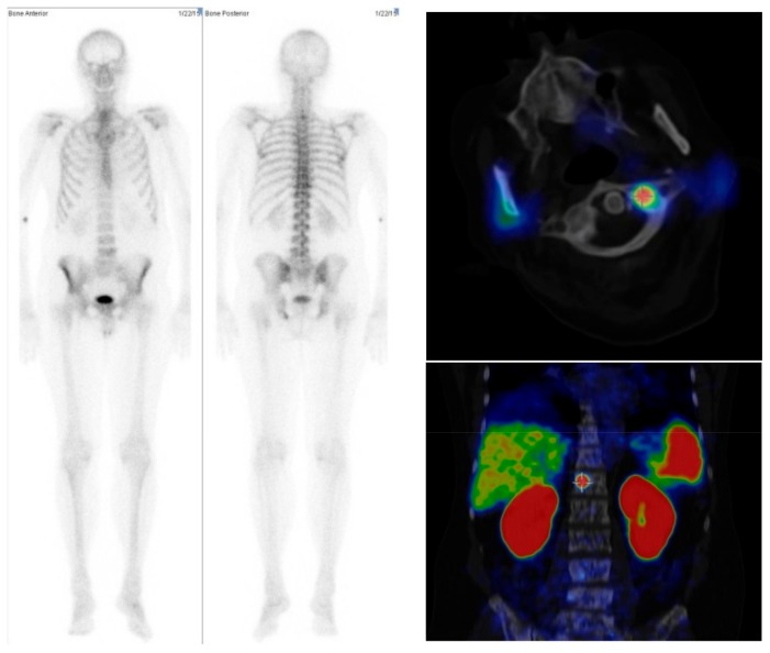
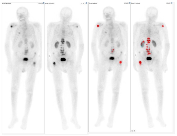
In the bone scans, visual and semiautomatic lesion detection showed similar results with an average of 19.4 and 17.8 detected bone lesion per patient. However, in PSMA-PET/CT, on average double the numbers of lesions (40.0) were detected. The largest differences were found in the thorax and pelvis, which can be explained by the advantages of tomographic imaging. Bland-Altman analysis showed greater differences in patients with large numbers of bone metastases.

CONCLUSION

No significant difference was found when using semiautomatic analysis compared to visual reading for bone scans. Fewer bone metastases were detected in bone scans than in PSMA-PET/CT. However, in none of our patients would the difference have led to clinical consequences. Therefore, it seems that for patients undergoing PSMA-PET/CT, there is no need to perform additional bone scans if the appropriate PET/CT protocols are applied.

摘要

目的

骨闪烁扫描是前列腺癌患者骨转移的参考标准。然而,前列腺特异性膜抗原(PSMA)配体中使用的新型放射性示踪剂使得PET/CT作为诊断工具的重要性日益增加。我们研究的目的是探讨骨扫描与PSMA-PET/CT在检测前列腺癌骨转移方面的差异。

方法

30例前列腺癌骨转移患者在平均21天内接受了Tc-MDP骨扫描和Ga-PSMA-PET/CT检查。根据病变数量对骨扫描进行视觉分析,并使用Exini Diagnostics公司的ExiniBONE软件包进行分析。对PET/CT数据进行视觉分析。比较了不同方法对全患者和不同区域检测到的病变数量。此外,将结果与血清前列腺特异性抗原(PSA)、碱性磷酸酶(ALP)、骨碱性磷酸酶(bALP)、促胃泌素释放肽(pGRP)以及东部肿瘤协作组(ECOG)的体能状态进行了比较。

结果

在骨扫描中,视觉和半自动病变检测结果相似,每位患者平均检测到19.4个和17.8个骨病变。然而,在PSMA-PET/CT中,平均检测到的病变数量(40.0个)是其两倍。在胸部和骨盆中发现的差异最大,这可以通过断层成像的优势来解释。Bland-Altman分析显示,骨转移数量较多的患者差异更大。

结论

与骨扫描的视觉读数相比,半自动分析未发现显著差异。骨扫描检测到的骨转移比PSMA-PET/CT少。然而,在我们的患者中,这种差异均未导致临床后果。因此,对于接受PSMA-PET/CT检查的患者,如果应用了适当的PET/CT方案,似乎无需进行额外的骨扫描。

https://cdn.ncbi.nlm.nih.gov/pmc/blobs/fdca/5620612/a244229b5c50/pharmaceuticals-10-00068-g004.jpg
https://cdn.ncbi.nlm.nih.gov/pmc/blobs/fdca/5620612/bbd0b5d84881/pharmaceuticals-10-00068-g001.jpg
https://cdn.ncbi.nlm.nih.gov/pmc/blobs/fdca/5620612/16c2806f3e51/pharmaceuticals-10-00068-g002.jpg
https://cdn.ncbi.nlm.nih.gov/pmc/blobs/fdca/5620612/6834e5517166/pharmaceuticals-10-00068-g003.jpg
https://cdn.ncbi.nlm.nih.gov/pmc/blobs/fdca/5620612/a244229b5c50/pharmaceuticals-10-00068-g004.jpg

相似文献

1
Assessment of Bone Metastases in Patients with Prostate Cancer-A Comparison between Tc-Bone-Scintigraphy and [Ga]Ga-PSMA PET/CT.前列腺癌患者骨转移的评估——锝骨闪烁显像与[镓]镓-PSMA PET/CT的比较
Pharmaceuticals (Basel). 2017 Jul 31;10(3):68. doi: 10.3390/ph10030068.
2
More advantages in detecting bone and soft tissue metastases from prostate cancer using F-PSMA PET/CT.使用F-PSMA PET/CT检测前列腺癌骨和软组织转移方面有更多优势。
Hell J Nucl Med. 2019 Jan-Apr;22(1):6-9. doi: 10.1967/s002449910952. Epub 2019 Mar 7.
3
A prospective intra-individual comparison of [Ga]Ga-PSMA-11 PET/CT, [Ga]Ga-NODAGA PET/CT, and [Tc]Tc-MDP bone scintigraphy for radionuclide imaging of prostate cancer skeletal metastases.一种前瞻性个体内比较研究:用于前列腺癌骨转移放射性核素成像的[Ga]Ga-PSMA-11 PET/CT、[Ga]Ga-NODAGA PET/CT 和[Tc]Tc-MDP 骨闪烁显像。
Eur J Nucl Med Mol Imaging. 2021 Jan;48(1):134-142. doi: 10.1007/s00259-020-04867-y. Epub 2020 May 18.
4
Head-To-Head Comparison of Ga-PSMA-11 PET/CT and Tc-MDP Bone Scintigraphy for the Detection of Bone Metastases in Patients With Prostate Cancer: A Meta-Analysis.镓-PSMA-11 PET/CT 与 Tc-MDP 骨显像用于前列腺癌患者骨转移检测的头对头比较:一项荟萃分析。
AJR Am J Roentgenol. 2022 Sep;219(3):386-395. doi: 10.2214/AJR.21.27323. Epub 2022 Apr 20.
5
The diagnostic accuracy of Ga-PSMA PET/CT versus Tc-MDP bone scintigraphy for identifying bone metastases in persons with prostate cancer: A systematic review.镓-前列腺特异性膜抗原正电子发射断层扫描/计算机断层扫描(Ga-PSMA PET/CT)与锝-亚甲基二膦酸盐骨闪烁显像(Tc-MDP)在前列腺癌患者骨转移诊断中的准确性:一项系统评价。
J Med Imaging Radiat Sci. 2023 Sep;54(3):545-555. doi: 10.1016/j.jmir.2023.04.005. Epub 2023 May 20.
6
Prostate-specific membrane antigen PET versus [Tc]Tc-MDP bone scan for diagnosing bone metastasis in prostate cancer: a head-to-head comparative meta-analysis.前列腺特异性膜抗原正电子发射断层扫描与[锝]锝-亚甲基二膦酸盐骨扫描在诊断前列腺癌骨转移中的应用:一项直接比较的Meta分析
Front Med (Lausanne). 2024 Sep 25;11:1451565. doi: 10.3389/fmed.2024.1451565. eCollection 2024.
7
Comparison of hybrid Ga-PSMA-PET/CT and Tc-DPD-SPECT/CT for the detection of bone metastases in prostate cancer patients: Additional value of morphologic information from low dose CT.镓-PSMA-PET/CT 与 Tc-DPD-SPECT/CT 比较在前列腺癌患者骨转移检测中的应用:低剂量 CT 形态学信息的附加价值。
Eur Radiol. 2018 Feb;28(2):610-619. doi: 10.1007/s00330-017-4994-6. Epub 2017 Aug 4.
8
Intraindividual Comparison of Tc-Methylene Diphosphonate and Prostate-Specific Membrane Antigen Ligand Tc-MIP-1427 in Patients with Osseous Metastasized Prostate Cancer.Tc-亚甲基二膦酸盐与前列腺特异性膜抗原配体 Tc-MIP-1427 用于骨转移前列腺癌患者的个体内比较。
J Nucl Med. 2018 Sep;59(9):1373-1379. doi: 10.2967/jnumed.117.200220. Epub 2018 Jan 25.
9
Added value of Ga-PSMA PET/CT for the detection of bone metastases in patients with newly diagnosed prostate cancer and a previous Tc bone scintigraphy.镓-前列腺特异性膜抗原正电子发射断层扫描/计算机断层扫描(Ga-PSMA PET/CT)在新诊断前列腺癌且既往行锝骨闪烁显像患者骨转移检测中的附加价值
EJNMMI Res. 2020 Apr 8;10(1):31. doi: 10.1186/s13550-020-00618-0.
10
Direct comparison of Tc-PSMA SPECT/CT and Ga-PSMA PET/CT in patients with prostate cancer.前列腺癌患者中锝-PSMA单光子发射计算机断层扫描/计算机断层扫描(Tc-PSMA SPECT/CT)与镓-PSMA正电子发射断层扫描/计算机断层扫描(Ga-PSMA PET/CT)的直接比较。
Asia Ocean J Nucl Med Biol. 2020 Winter;8(1):1-7. doi: 10.22038/aojnmb.2019.43943.1293.

引用本文的文献

1
Prostate-Specific Membrane Antigen Positron Emission Tomography (PSMA-PET) in Initial Staging of Prostate Cancer Patients: The Beginning of a New Era.前列腺特异性膜抗原正电子发射断层扫描(PSMA-PET)在前列腺癌患者初始分期中的应用:新时代的开端。
Medicina (Kaunas). 2025 May 20;61(5):924. doi: 10.3390/medicina61050924.
2
Progress in Oligometastatic Prostate Cancer: Emerging Imaging Innovations and Therapeutic Approaches.寡转移前列腺癌的进展:新兴的影像创新与治疗方法
Cancers (Basel). 2024 Jan 24;16(3):507. doi: 10.3390/cancers16030507.
3
Underlying Features of Prostate Cancer-Statistics, Risk Factors, and Emerging Methods for Its Diagnosis.

本文引用的文献

1
Exploring New Multimodal Quantitative Imaging Indices for the Assessment of Osseous Tumor Burden in Prostate Cancer Using Ga-PSMA PET/CT.探讨使用 Ga-PSMA PET/CT 评估前列腺癌骨肿瘤负荷的新型多模态定量成像指标。
J Nucl Med. 2017 Oct;58(10):1632-1637. doi: 10.2967/jnumed.116.189050. Epub 2017 May 25.
2
Comparison of bone scintigraphy and Ga-PSMA PET for skeletal staging in prostate cancer.骨闪烁显像与镓-PSMA PET在前列腺癌骨骼分期中的比较
Eur J Nucl Med Mol Imaging. 2016 Nov;43(12):2114-2121. doi: 10.1007/s00259-016-3435-0. Epub 2016 Jun 12.
3
Molecular Imaging of Prostate Cancer.
前列腺癌的基本特征-统计数据、风险因素和新的诊断方法。
Curr Oncol. 2023 Feb 15;30(2):2300-2321. doi: 10.3390/curroncol30020178.
4
[Diagnosis and staging of metastatic hormone-sensitive prostate cancer].[转移性激素敏感性前列腺癌的诊断与分期]
Urologie. 2023 Apr;62(4):347-353. doi: 10.1007/s00120-023-02028-1. Epub 2023 Feb 3.
5
Head-to-head comparison of Tc-PSMA and Tc-MDP SPECT/CT in diagnosing prostate cancer bone metastasis: a prospective, comparative imaging trial.Tc-PSMA 与 Tc-MDP SPECT/CT 对头对头比较诊断前列腺癌骨转移:一项前瞻性、对比成像试验。
Sci Rep. 2022 Sep 26;12(1):15993. doi: 10.1038/s41598-022-20280-x.
6
Where Do We Stand in the Management of Oligometastatic Prostate Cancer? A Comprehensive Review.在寡转移前列腺癌的管理方面我们处于什么位置?一项全面综述。
Cancers (Basel). 2022 Apr 16;14(8):2017. doi: 10.3390/cancers14082017.
7
Emerging methods for prostate cancer imaging: evaluating cancer structure and metabolic alterations more clearly.新兴的前列腺癌成像方法:更清晰地评估癌症结构和代谢变化。
Mol Oncol. 2021 Oct;15(10):2565-2579. doi: 10.1002/1878-0261.13071. Epub 2021 Aug 30.
8
Phase 3 multicenter randomized trial of PSMA PET/CT prior to definitive radiation therapy for unfavorable intermediate-risk or high-risk prostate cancer [PSMA dRT]: study protocol.三期多中心随机临床试验,研究 PSMA PET/CT 在前瞻性放射性治疗中对于不利的中危或高危前列腺癌(PSMA dRT)的作用:研究方案。
BMC Cancer. 2021 May 7;21(1):512. doi: 10.1186/s12885-021-08026-w.
9
Volume matters and intensification is needed: emerging trends in the management of advanced prostate cancer.体积很重要,强化治疗不可或缺:晚期前列腺癌管理的新趋势
Drugs Context. 2021 Mar 15;10. doi: 10.7573/dic.2020-10-2. eCollection 2021.
10
Multimodal imaging for radiation therapy planning in patients with primary prostate cancer.原发性前列腺癌患者放射治疗计划的多模态成像
Phys Imaging Radiat Oncol. 2018 Nov 5;8:8-16. doi: 10.1016/j.phro.2018.10.001. eCollection 2018 Oct.
前列腺癌的分子成像
Radiographics. 2016 Jan-Feb;36(1):142-59. doi: 10.1148/rg.2016150059. Epub 2015 Nov 20.
4
Comparison of hybrid (68)Ga-PSMA PET/MRI and (68)Ga-PSMA PET/CT in the evaluation of lymph node and bone metastases of prostate cancer.(68)镓-前列腺特异性膜抗原正电子发射断层扫描/磁共振成像与(68)镓-前列腺特异性膜抗原正电子发射断层扫描/计算机断层扫描在评估前列腺癌淋巴结和骨转移中的比较
Eur J Nucl Med Mol Imaging. 2016 Jan;43(1):70-83. doi: 10.1007/s00259-015-3206-3. Epub 2015 Oct 28.
5
EXINI Quantitative Bone Scan Index: Expanded Utility for the Planar Radionuclide Bone Scan.EXINI定量骨扫描指数:平面放射性核素骨扫描的扩展应用
J Nucl Med. 2016 Jan;57(1):5-6. doi: 10.2967/jnumed.115.164137. Epub 2015 Oct 8.
6
Prospective Comparison of 99mTc-MDP Scintigraphy, Combined 18F-NaF and 18F-FDG PET/CT, and Whole-Body MRI in Patients with Breast and Prostate Cancer.99mTc-MDP 闪烁显像、联合 18F-NaF 和 18F-FDG PET/CT、全身 MRI 在乳腺癌和前列腺癌患者中的前瞻性比较。
J Nucl Med. 2015 Dec;56(12):1862-8. doi: 10.2967/jnumed.115.162610. Epub 2015 Sep 24.
7
Analytic Validation of the Automated Bone Scan Index as an Imaging Biomarker to Standardize Quantitative Changes in Bone Scans of Patients with Metastatic Prostate Cancer.自动化骨扫描指数的分析验证:作为一种成像生物标志物,用于标准化转移性前列腺癌患者骨扫描的定量变化。
J Nucl Med. 2016 Jan;57(1):41-5. doi: 10.2967/jnumed.115.160085. Epub 2015 Aug 27.
8
Evaluation of Hybrid ⁶⁸Ga-PSMA Ligand PET/CT in 248 Patients with Biochemical Recurrence After Radical Prostatectomy.⁶⁸Ga-PSMA配体杂交正电子发射断层显像/计算机断层扫描在248例前列腺癌根治术后生化复发患者中的评估
J Nucl Med. 2015 May;56(5):668-74. doi: 10.2967/jnumed.115.154153. Epub 2015 Mar 19.
9
(68)Ga-PSMA PET/MR with multimodality image analysis for primary prostate cancer.用于原发性前列腺癌的(68)镓-前列腺特异性膜抗原正电子发射断层扫描/磁共振成像及多模态图像分析
Abdom Imaging. 2015 Aug;40(6):1769-71. doi: 10.1007/s00261-014-0301-z.
10
EAU guidelines on prostate cancer. part 1: screening, diagnosis, and local treatment with curative intent-update 2013.EAU 前列腺癌指南。第 1 部分:筛查、诊断和以治愈为目的的局部治疗——2013 年更新。
Eur Urol. 2014 Jan;65(1):124-37. doi: 10.1016/j.eururo.2013.09.046. Epub 2013 Oct 6.