Institute of Integrative Biology, University of Liverpool, Crown Street, Liverpool L69 7ZB, U.K.
Institute of Infection and Global Health, University of Liverpool, Crown Street, Liverpool L69 7ZB, U.K.
Biochem J. 2020 Mar 27;477(6):1159-1178. doi: 10.1042/BCJ20190644.
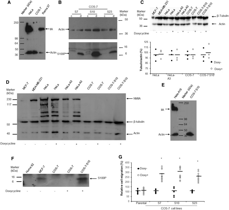
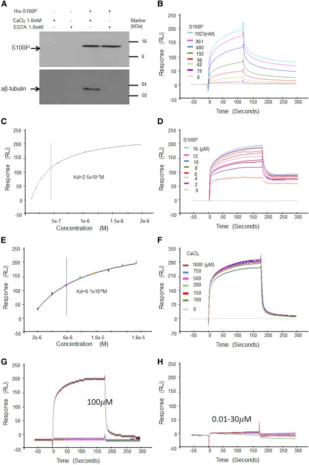
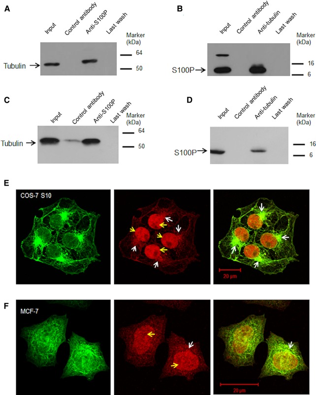
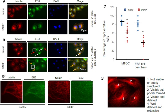
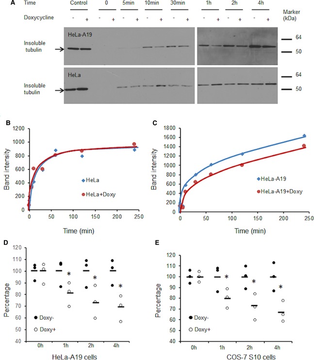
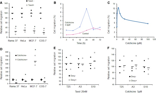
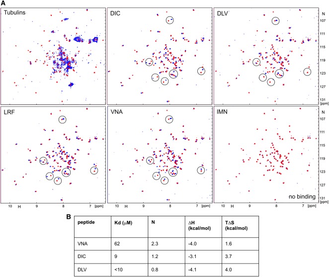
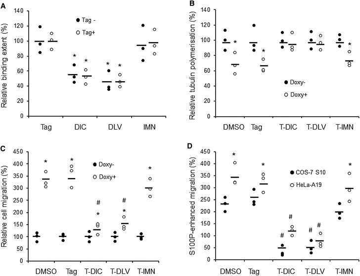
Overexpression of S100P promotes breast cancer metastasis in animals and elevated levels in primary breast cancers are associated with poor patient outcomes. S100P can differentially interact with nonmuscle myosin (NM) isoforms (IIA > IIC > IIB) leading to the redistribution of actomyosin filaments to enhance cell migration. Using COS-7 cells which do not naturally express NMIIA, S100P is now shown to interact directly with α,β-tubulin in vitro and in vivo with an equilibrium Kd of 2-3 × 10⁻⁷ M. The overexpressed S100P is located mainly in nuclei and microtubule organising centres (MTOC) and it significantly reduces their number, slows down tubulin polymerisation and enhances cell migration in S100P-induced COS-7 or HeLa cells. It fails, however, to significantly reduce cell adhesion, in contrast with NMIIA-containing S100P-inducible HeLa cells. When taxol is used to stabilise MTs or colchicine to dissociate MTs, S100P's stimulation of migration is abolished. Affinity-chromatography of tryptic digests of α and β-tubulin on S100P-bound beads identifies multiple S100P-binding sites consistent with S100P binding to all four half molecules in gel-overlay assays. When screened by NMR and ITC for interacting with S100P, four chemically synthesised peptides show interactions with low micromolar dissociation constants. The two highest affinity peptides significantly inhibit binding of S100P to α,β-tubulin and, when tagged for cellular entry, also inhibit S100P-induced reduction in tubulin polymerisation and S100P-enhancement of COS-7 or HeLa cell migration. A third peptide incapable of interacting with S100P also fails in this respect. Thus S100P can interact directly with two different cytoskeletal filaments to independently enhance cell migration, the most important step in the metastatic cascade.
S100P 的过表达促进了动物乳腺癌的转移,而原发性乳腺癌中 S100P 水平的升高与患者预后不良有关。S100P 可以与非肌肉肌球蛋白(NM)同工型(IIA> IIC> IIB)不同地相互作用,导致肌动球蛋白丝的重新分布,从而增强细胞迁移。使用不自然表达 NMIIA 的 COS-7 细胞,现在已经证明 S100P 可以在体外和体内与α、β-微管蛋白直接相互作用,平衡 Kd 为 2-3×10⁻⁷ M。过表达的 S100P 主要位于细胞核和微管组织中心(MTOC),它显著减少了它们的数量,减缓了微管蛋白聚合,并增强了 S100P 诱导的 COS-7 或 HeLa 细胞的迁移。然而,它不能显著降低细胞黏附力,与含有 NMIIA 的 S100P 诱导的 HeLa 细胞形成对比。当紫杉醇用于稳定 MT 或秋水仙素用于解离 MT 时,S100P 对迁移的刺激作用被消除。S100P 结合珠上的胰蛋白酶消化物的亲和层析鉴定出多个与 S100P 结合的位点,这与凝胶覆盖实验中 S100P 与所有四个半分子结合一致。通过 NMR 和 ITC 筛选与 S100P 相互作用的物质,四种化学合成的肽显示出与低微摩尔解离常数的相互作用。两个亲和力最高的肽显著抑制 S100P 与α、β-微管蛋白的结合,并且当标记为细胞进入时,也抑制 S100P 诱导的微管蛋白聚合减少和 S100P 增强的 COS-7 或 HeLa 细胞迁移。不能与 S100P 相互作用的第三种肽在这方面也失败了。因此,S100P 可以直接与两种不同的细胞骨架丝相互作用,独立地增强细胞迁移,这是转移级联中最重要的步骤。