Cardiology Department Hospital Clínico Universitario de Valencia Universidad de Valencia INCLIVA Valencia Spain.
CIBER Cardiovascular Universitat Jaume I Castellón Spain.
J Am Heart Assoc. 2020 Feb 18;9(4):e014254. doi: 10.1161/JAHA.119.014254. Epub 2020 Feb 13.
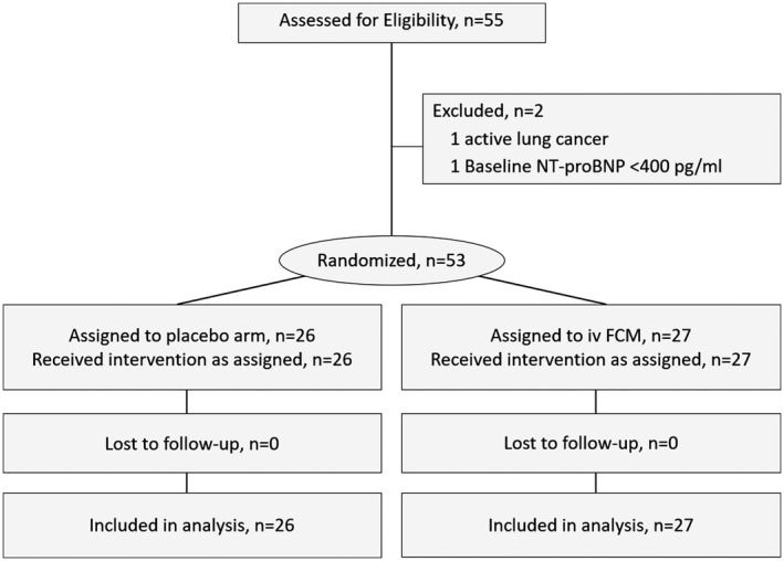
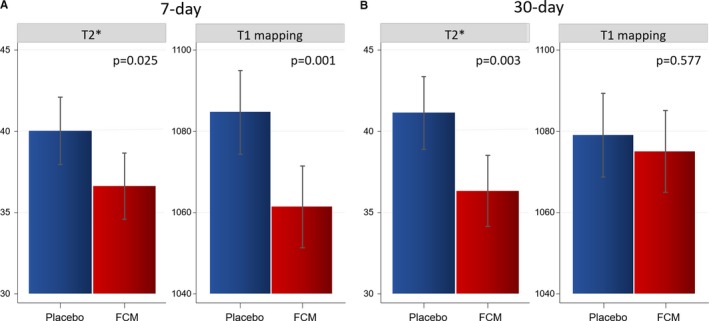
Background Intravenous ferric carboxymaltose (FCM) improves symptoms, functional capacity, and quality of life in heart failure and iron deficiency. The mechanisms underlying these effects are not fully understood. The aim of this study was to examine changes in myocardial iron content after FCM administration in patients with heart failure and iron deficiency using cardiac magnetic resonance. Methods and Results Fifty-three stable heart failure and iron deficiency patients were randomly assigned 1:1 to receive intravenous FCM or placebo in a multicenter, double-blind study. T2* and T1 mapping cardiac magnetic resonance sequences, noninvasive surrogates of intramyocardial iron, were evaluated before and 7 and 30 days after randomization using linear mixed regression analysis. Results are presented as least-square means with 95% CI. The primary end point was the change in T2* and T1 mapping at 7 and 30 days. Median age was 73 (65-78) years, with N-terminal pro-B-type natriuretic peptide, ferritin, and transferrin saturation medians of 1690 pg/mL (1010-2828), 63 ng/mL (22-114), and 15.7% (11.0-19.2), respectively. Baseline T2* and T1 mapping values did not significantly differ across treatment arms. On day 7, both T2* and T1 mapping (ms) were significantly lower in the FCM arm (36.6 [34.6-38.7] versus 40 [38-42.1], =0.025; 1061 [1051-1072] versus 1085 [1074-1095], =0.001, respectively). A similar reduction was found at 30 days for T2* (36.3 [34.1-38.5] versus 41.1 [38.9-43.4], =0.003), but not for T1 mapping (1075 [1065-1085] versus 1079 [1069-1089], =0.577). Conclusions In patients with heart failure and iron deficiency, FCM administration was associated with changes in the T2* and T1 mapping cardiac magnetic resonance sequences, indicative of myocardial iron repletion. Clinical Trial Registration URL: http://www.clinicaltrials.gov. Unique identifier: NCT03398681.
静脉注射羧基麦芽糖铁(FCM)可改善心力衰竭和缺铁患者的症状、功能能力和生活质量。但其作用机制尚未完全阐明。本研究旨在使用心脏磁共振评估心力衰竭和缺铁患者接受 FCM 治疗后心肌铁含量的变化。
53 例稳定的心力衰竭和缺铁患者以 1:1 的比例随机接受静脉注射 FCM 或安慰剂,在一项多中心、双盲研究中进行。使用线性混合回归分析,在随机分组前及 7 天和 30 天时评估 T2和 T1 映射心脏磁共振序列,这是非侵入性心肌内铁的替代指标。结果以最小二乘均数(95%CI)表示。主要终点为 7 天和 30 天时 T2和 T1 映射的变化。中位年龄为 73(65-78)岁,N 端脑钠肽前体、铁蛋白和转铁蛋白饱和度中位数分别为 1690pg/mL(1010-2828)、63ng/mL(22-114)和 15.7%(11.0-19.2)。治疗组之间的基线 T2和 T1 映射值无显著差异。在第 7 天,FCM 组的 T2和 T1 映射(ms)均显著降低(36.6[34.6-38.7] 与 40[38-42.1],=0.025;1061[1051-1072] 与 1085[1074-1095],=0.001)。在 30 天时,T2*也观察到类似的降低(36.3[34.1-38.5] 与 41.1[38.9-43.4],=0.003),但 T1 映射未见降低(1075[1065-1085] 与 1079[1069-1089],=0.577)。
在心力衰竭和缺铁患者中,FCM 给药与 T2*和 T1 映射心脏磁共振序列的变化相关,表明心肌铁补充。