• 文献检索
  • 文档翻译
  • 深度研究
  • 学术资讯
  • Suppr Zotero 插件Zotero 插件
  • 邀请有礼
  • 套餐&价格
  • 历史记录
应用&插件
Suppr Zotero 插件Zotero 插件浏览器插件Mac 客户端Windows 客户端微信小程序
定价
高级版会员购买积分包购买API积分包
服务
文献检索文档翻译深度研究API 文档MCP 服务
关于我们
关于 Suppr公司介绍联系我们用户协议隐私条款
关注我们

Suppr 超能文献

核心技术专利:CN118964589B侵权必究
粤ICP备2023148730 号-1Suppr @ 2026

文献检索

告别复杂PubMed语法,用中文像聊天一样搜索,搜遍4000万医学文献。AI智能推荐,让科研检索更轻松。

立即免费搜索

文件翻译

保留排版,准确专业,支持PDF/Word/PPT等文件格式,支持 12+语言互译。

免费翻译文档

深度研究

AI帮你快速写综述,25分钟生成高质量综述,智能提取关键信息,辅助科研写作。

立即免费体验

3D 生物打印软骨细胞至骨关节炎软骨胫骨病变后 2 型胶原 B 的诱导。

Collagen 2A Type B Induction after 3D Bioprinting Chondrocytes into Osteoarthritic Chondral Tibial Lesion.

机构信息

Department of Orthopaedics, Institute of Clinical Sciences, the Sahlgrenska Academy at the University of Gothenburg, Gothenburg, Sweden.

Sahlgrenska University Hospital, Mölndal, Sweden.

出版信息

Cartilage. 2021 Dec;13(2_suppl):1755S-1769S. doi: 10.1177/1947603520903788. Epub 2020 Feb 18.

DOI:10.1177/1947603520903788
PMID:32070108
原文链接:https://pmc.ncbi.nlm.nih.gov/articles/PMC8721610/
Abstract

OBJECTIVE

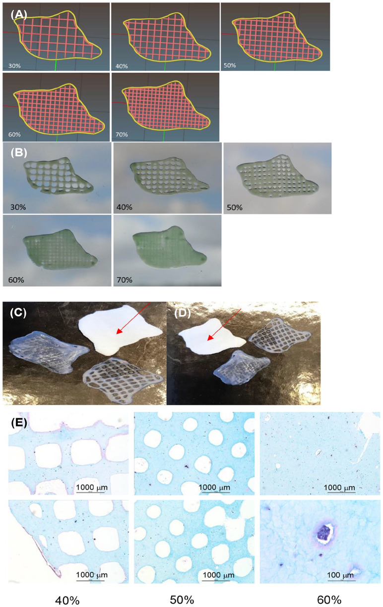
Large cartilage defects and osteoarthritis (OA) cause cartilage loss and remain a therapeutic challenge. Three-dimensional (3D) bioprinting with autologous cells using a computer-aided design (CAD) model generated from 3D imaging has the potential to reconstruct patient-specific features that match an articular joint lesion.

DESIGN

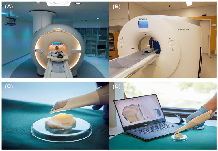
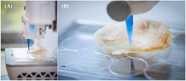
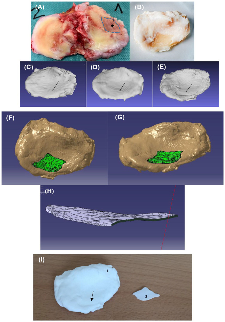
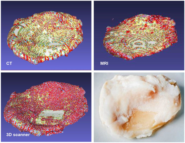
To scan a human OA tibial plateau with a cartilage defect, retrieved after total knee arthroplasty, following clinical imaging techniques were used: (1) computed tomography (CT), (2) magnetic resonance imaging (MRI), and (3) a 3D scanner. From such a scan, a CAD file was obtained to generate G-code to control 3D bioprinting directly into the tibial plateau lesion.

RESULTS

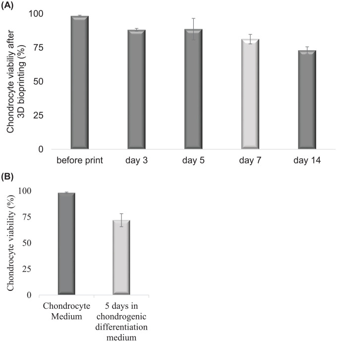
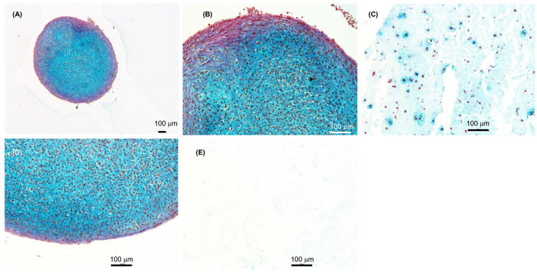
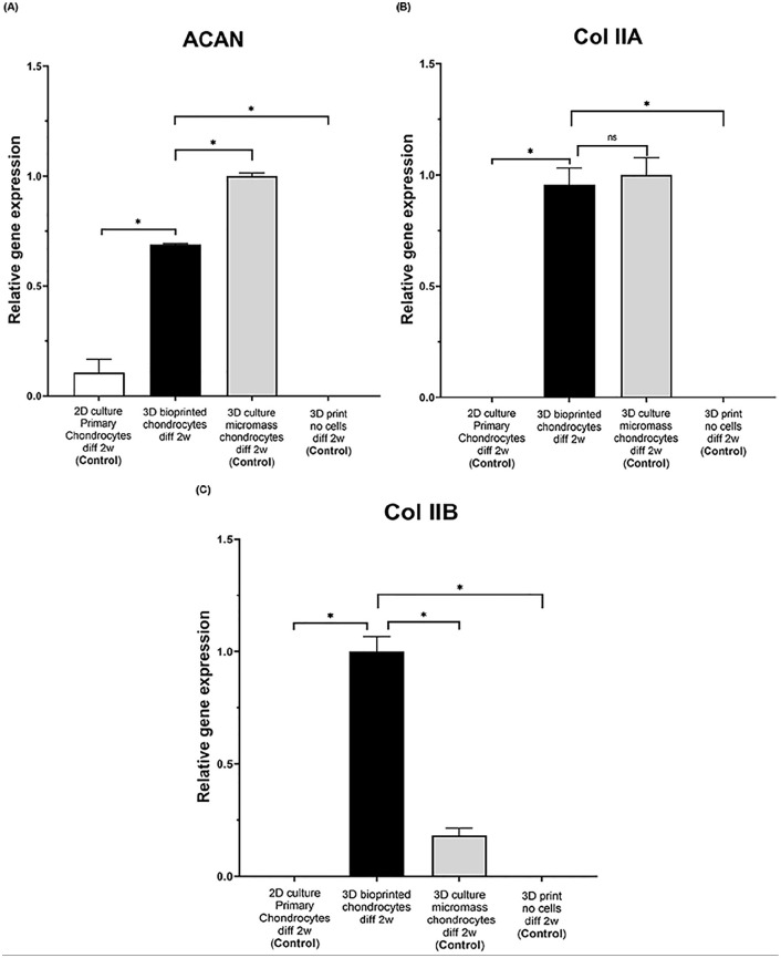
Highest resolution was obtained using the 3D scanner (2.77 times more points/mm than CT), and of the 3 devices tested, only the 3D scanner was able to detect the actual OA defect area. Human chondrocytes included in 3D bioprinted constructs produced extracellular matrix and formed cartilage tissue fragments after 2 weeks of differentiation and high levels of a mature splice version of collagen type II (Col IIA type B), characteristic of native articular cartilage and aggrecan (ACAN). Chondrocytes had a mean viability of 81% in prints after day 5 of differentiation toward cartilage and similar viability was detected in control 3D pellet differentiation of chondrocytes (mean viability 72%).

CONCLUSION

Articular cartilage can be formed in 3D bioprints. Thus, this 3D bioprinting system with chondrocytes simulating a patient-specific 3D model provides an attractive strategy for future treatments of cartilage defects or early OA.

摘要

目的

大的软骨缺损和骨关节炎(OA)导致软骨丢失,仍然是一个治疗挑战。使用源自 3D 成像的计算机辅助设计(CAD)模型的自体细胞 3D 生物打印具有重建与关节病变匹配的患者特定特征的潜力。

设计

为了扫描经全膝关节置换术切除的人类 OA 胫骨平台软骨缺损,采用以下临床成像技术进行扫描:(1)计算机断层扫描(CT),(2)磁共振成像(MRI)和(3)3D 扫描仪。从这样的扫描中,获得 CAD 文件以生成 G 代码来直接控制 3D 生物打印到胫骨平台病变中。

结果

使用 3D 扫描仪获得了最高分辨率(比 CT 多 2.77 倍的点/mm),在测试的 3 种设备中,只有 3D 扫描仪能够检测到实际的 OA 缺陷区域。包含在 3D 生物打印构建体中的人软骨细胞在分化后的 2 周内产生细胞外基质并形成软骨组织碎片,并且具有高比例的成熟拼接型 II 型胶原(Col IIA 型 B),这是天然关节软骨和聚集蛋白聚糖(ACAN)的特征。在向软骨分化的第 5 天,打印中的软骨细胞平均存活率为 81%,在对照 3D 软骨细胞球分化中检测到相似的存活率(平均存活率为 72%)。

结论

可以在 3D 生物打印中形成关节软骨。因此,这种具有模拟患者特定 3D 模型的软骨细胞的 3D 生物打印系统为将来治疗软骨缺损或早期 OA 提供了有吸引力的策略。

https://cdn.ncbi.nlm.nih.gov/pmc/blobs/83b2/8804735/3c22dc736a0f/10.1177_1947603520903788-fig8.jpg
https://cdn.ncbi.nlm.nih.gov/pmc/blobs/83b2/8804735/28140fcf07cf/10.1177_1947603520903788-fig1.jpg
https://cdn.ncbi.nlm.nih.gov/pmc/blobs/83b2/8804735/53c09c49c07a/10.1177_1947603520903788-fig2.jpg
https://cdn.ncbi.nlm.nih.gov/pmc/blobs/83b2/8804735/7eec51b3e098/10.1177_1947603520903788-fig3.jpg
https://cdn.ncbi.nlm.nih.gov/pmc/blobs/83b2/8804735/1ecddd6d2c7e/10.1177_1947603520903788-fig4.jpg
https://cdn.ncbi.nlm.nih.gov/pmc/blobs/83b2/8804735/1c251392835e/10.1177_1947603520903788-fig5.jpg
https://cdn.ncbi.nlm.nih.gov/pmc/blobs/83b2/8804735/6512363c1df4/10.1177_1947603520903788-fig6.jpg
https://cdn.ncbi.nlm.nih.gov/pmc/blobs/83b2/8804735/6def6f9f8b0d/10.1177_1947603520903788-fig7.jpg
https://cdn.ncbi.nlm.nih.gov/pmc/blobs/83b2/8804735/3c22dc736a0f/10.1177_1947603520903788-fig8.jpg

相似文献

1
Collagen 2A Type B Induction after 3D Bioprinting Chondrocytes into Osteoarthritic Chondral Tibial Lesion.3D 生物打印软骨细胞至骨关节炎软骨胫骨病变后 2 型胶原 B 的诱导。
Cartilage. 2021 Dec;13(2_suppl):1755S-1769S. doi: 10.1177/1947603520903788. Epub 2020 Feb 18.
2
Expression of collagen and aggrecan genes in normal and osteoarthritic murine knee joints.正常和骨关节炎小鼠膝关节中胶原蛋白和聚集蛋白聚糖基因的表达
Osteoarthritis Cartilage. 2002 Jan;10(1):51-61. doi: 10.1053/joca.2001.0481.
3
Assessing the advantages of 3D bioprinting and 3D spheroids in deciphering the osteoarthritis healing mechanism using human chondrocytes and polarized macrophages.评估 3D 生物打印和 3D 球体在利用人软骨细胞和极化巨噬细胞解析骨关节炎愈合机制方面的优势。
Biomed Mater. 2024 Jan 19;19(2). doi: 10.1088/1748-605X/ad1d18.
4
Multi-material 3D bioprinting of porous constructs for cartilage regeneration.多材料 3D 生物打印多孔结构用于软骨再生。
Mater Sci Eng C Mater Biol Appl. 2020 Apr;109:110578. doi: 10.1016/j.msec.2019.110578. Epub 2019 Dec 20.
5
Regional differences of tibial and femoral cartilage in the chondrocyte gene expression, immunhistochemistry and composite in different stages of osteoarthritis.骨关节炎不同阶段胫骨和股骨软骨在软骨细胞基因表达、免疫组织化学及复合物方面的区域差异。
Tissue Cell. 2017 Apr;49(2 Pt B):249-256. doi: 10.1016/j.tice.2017.02.004. Epub 2017 Feb 24.
6
Varying development of femoral and tibial subchondral bone tissue and their interaction with articular cartilage during progressing osteoarthritis.在进展性骨关节炎中,股骨和胫骨的软骨下骨组织的不同发育及其与关节软骨的相互作用。
Arch Orthop Trauma Surg. 2020 Dec;140(12):1919-1930. doi: 10.1007/s00402-020-03480-w. Epub 2020 May 30.
7
Scaffold Free Microtissue Formation for Enhanced Cartilage Repair.无支架微组织构建促进软骨修复。
Ann Biomed Eng. 2020 Jan;48(1):298-311. doi: 10.1007/s10439-019-02348-4. Epub 2019 Aug 26.
8
A 3D cartilage - inflammatory cell culture system for the modeling of human osteoarthritis.一种用于建模人类骨关节炎的 3D 软骨-炎性细胞培养系统。
Biomaterials. 2011 Aug;32(24):5581-9. doi: 10.1016/j.biomaterials.2011.04.028. Epub 2011 May 12.
9
3D bioprinting of photo-crosslinkable silk methacrylate (SilMA)-polyethylene glycol diacrylate (PEGDA) bioink for cartilage tissue engineering.用于软骨组织工程的光交联甲基丙烯酸丝素酯(SilMA)-聚乙二醇二丙烯酸酯(PEGDA)生物墨水的3D生物打印
J Biomed Mater Res A. 2022 Apr;110(4):884-898. doi: 10.1002/jbm.a.37336. Epub 2021 Dec 16.
10
Homeostasis of the extracellular matrix of normal and osteoarthritic human articular cartilage chondrocytes in vitro.体外培养的正常及骨关节炎人类关节软骨细胞细胞外基质的稳态
Osteoarthritis Cartilage. 2003 Nov;11(11):801-9. doi: 10.1016/s1063-4584(03)00168-7.

引用本文的文献

1
Targeting Chondrocyte Hypertrophy as Strategies for the Treatment of Osteoarthritis.以软骨细胞肥大作为骨关节炎治疗策略
Bioengineering (Basel). 2025 Jan 15;12(1):77. doi: 10.3390/bioengineering12010077.
2
3D-printed constructs deliver bioactive cargos to expedite cartilage regeneration.3D打印构建体递送生物活性物质以加速软骨再生。
J Pharm Anal. 2024 Dec;14(12):100925. doi: 10.1016/j.jpha.2023.12.015. Epub 2023 Dec 21.
3
A robotic arm with open-source reconstructive workflow for bioprinting of patient-specific scaffolds.

本文引用的文献

1
3D Bioprinting: New Directions in Articular Cartilage Tissue Engineering.3D生物打印:关节软骨组织工程的新方向。
ACS Biomater Sci Eng. 2017 Nov 13;3(11):2657-2668. doi: 10.1021/acsbiomaterials.6b00587. Epub 2017 Feb 8.
2
Chondrocytes and stem cells in 3D-bioprinted structures create human cartilage in vivo.3D生物打印结构中的软骨细胞和干细胞可在体内生成人体软骨。
PLoS One. 2017 Dec 13;12(12):e0189428. doi: 10.1371/journal.pone.0189428. eCollection 2017.
3
In situ repair of bone and cartilage defects using 3D scanning and 3D printing.
一种具有开源重建工作流程的机械臂,用于生物打印患者特异性支架。
Appl Phys Rev. 2024 Dec;11(4):041402. doi: 10.1063/5.0197123.
4
Application of 3D bioprinting technology apply to assessing Dangguiniantongtang (DGNT) decoctions in arthritis.3D生物打印技术在评估当归拈痛汤治疗关节炎中的应用。
Chin Med. 2024 Jul 8;19(1):96. doi: 10.1186/s13020-024-00948-4.
5
Bioprinting-Enabled Biomaterials: A Cutting-Edge Strategy for Future Osteoarthritis Therapy.生物打印技术赋能的生物材料:未来骨关节炎治疗的前沿策略。
Int J Nanomedicine. 2023 Nov 1;18:6213-6232. doi: 10.2147/IJN.S432468. eCollection 2023.
6
bioprinting: Broadening the therapeutic horizon for tissue injuries.生物打印:拓展组织损伤治疗的视野
Bioact Mater. 2023 Feb 6;25:201-222. doi: 10.1016/j.bioactmat.2023.01.018. eCollection 2023 Jul.
7
Portable hand-held bioprinters promote in situ tissue regeneration.便携式手持生物打印机促进原位组织再生。
Bioeng Transl Med. 2022 Mar 10;7(3):e10307. doi: 10.1002/btm2.10307. eCollection 2022 Sep.
8
Systematic review on the application of 3D-bioprinting technology in orthoregeneration: current achievements and open challenges.3D生物打印技术在骨再生中的应用系统综述:当前成果与开放挑战
J Exp Orthop. 2022 Sep 19;9(1):95. doi: 10.1186/s40634-022-00518-3.
9
Photo-Crosslinkable Hydrogels for 3D Bioprinting in the Repair of Osteochondral Defects: A Review of Present Applications and Future Perspectives.用于修复骨软骨缺损的3D生物打印的光交联水凝胶:当前应用及未来展望综述
Micromachines (Basel). 2022 Jun 29;13(7):1038. doi: 10.3390/mi13071038.
10
In situ bioprinting: intraoperative implementation of regenerative medicine.原位生物打印:再生医学的术中实施。
Trends Biotechnol. 2022 Oct;40(10):1229-1247. doi: 10.1016/j.tibtech.2022.03.009. Epub 2022 Apr 25.
利用三维扫描和三维打印原位修复骨和软骨缺损。
Sci Rep. 2017 Aug 25;7(1):9416. doi: 10.1038/s41598-017-10060-3.
4
The bio in the ink: cartilage regeneration with bioprintable hydrogels and articular cartilage-derived progenitor cells.墨水中的生物:可生物打印水凝胶和关节软骨衍生祖细胞的软骨再生。
Acta Biomater. 2017 Oct 1;61:41-53. doi: 10.1016/j.actbio.2017.08.005. Epub 2017 Aug 4.
5
Cartilage Tissue Engineering by the 3D Bioprinting of iPS Cells in a Nanocellulose/Alginate Bioink.利用纳米纤维素/藻酸盐生物墨水 3D 打印 iPS 细胞构建软骨组织工程
Sci Rep. 2017 Apr 6;7(1):658. doi: 10.1038/s41598-017-00690-y.
6
In Vivo Chondrogenesis in 3D Bioprinted Human Cell-laden Hydrogel Constructs.3D生物打印含人细胞水凝胶构建体中的体内软骨生成
Plast Reconstr Surg Glob Open. 2017 Feb 15;5(2):e1227. doi: 10.1097/GOX.0000000000001227. eCollection 2017 Feb.
7
Cell-laden hydrogels for osteochondral and cartilage tissue engineering.用于骨软骨和软骨组织工程的载细胞水凝胶。
Acta Biomater. 2017 Jul 15;57:1-25. doi: 10.1016/j.actbio.2017.01.036. Epub 2017 Jan 11.
8
Enhanced articular cartilage by human mesenchymal stem cells in enzymatically mediated transiently RGDS-functionalized collagen-mimetic hydrogels.人骨髓间充质干细胞在酶介导的瞬时RGDS功能化胶原模拟水凝胶中增强关节软骨。
Acta Biomater. 2017 Mar 15;51:75-88. doi: 10.1016/j.actbio.2017.01.028. Epub 2017 Jan 10.
9
Alginate Sulfate-Nanocellulose Bioinks for Cartilage Bioprinting Applications.用于软骨生物打印应用的硫酸藻酸盐-纳米纤维素生物墨水
Ann Biomed Eng. 2017 Jan;45(1):210-223. doi: 10.1007/s10439-016-1704-5. Epub 2016 Aug 8.
10
3D Bioprinting Human Chondrocytes with Nanocellulose-Alginate Bioink for Cartilage Tissue Engineering Applications.用于软骨组织工程应用的、采用纳米纤维素-藻酸盐生物墨水3D生物打印人软骨细胞
Biomacromolecules. 2015 May 11;16(5):1489-96. doi: 10.1021/acs.biomac.5b00188. Epub 2015 Apr 7.