Division of Anatomy, Department of Molecular Medicine, Institute of Basic Medical Sciences, University of Oslo, Blindern, Post box 1105, 0317, Oslo, Norway.
Department of Pathology, Oslo University Hospital, Oslo, Norway.
Brain Struct Funct. 2020 Mar;225(2):805-816. doi: 10.1007/s00429-020-02036-3. Epub 2020 Feb 18.
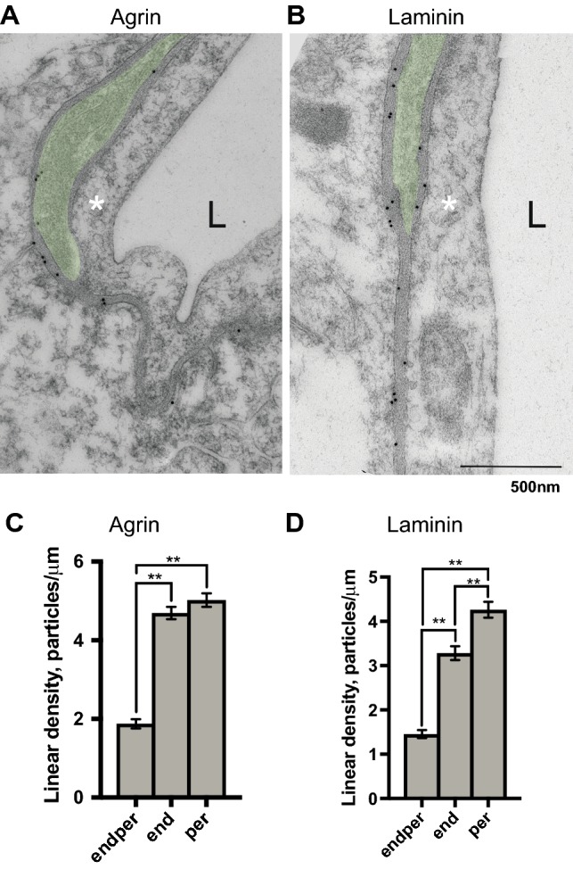
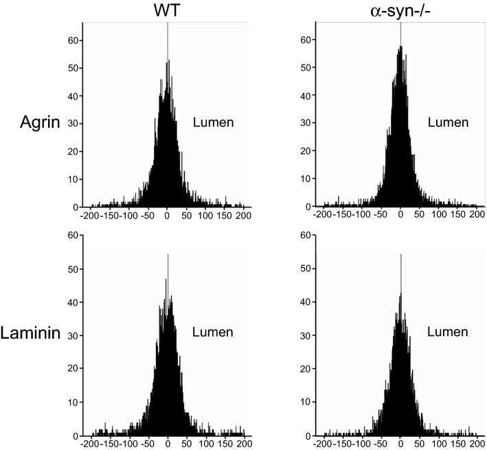
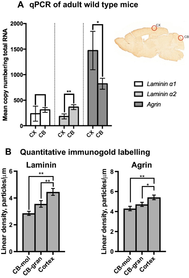
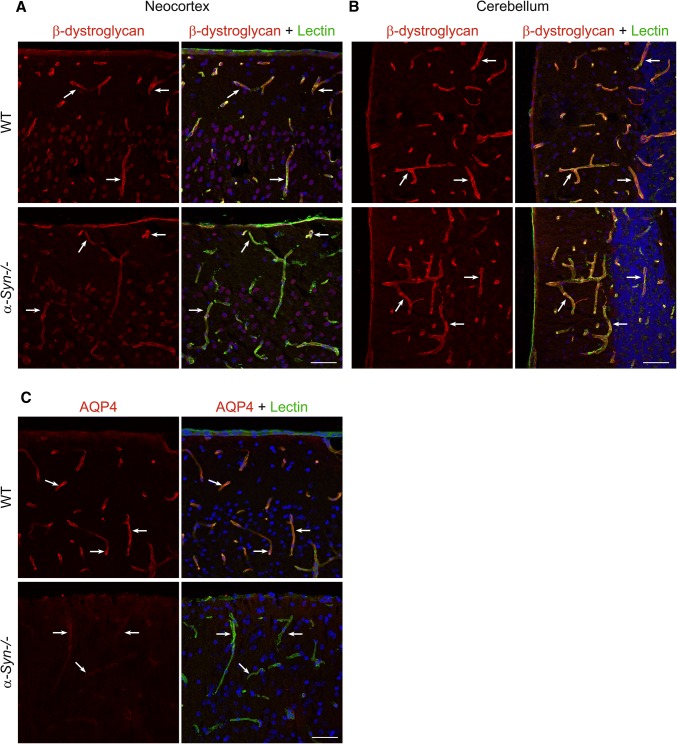
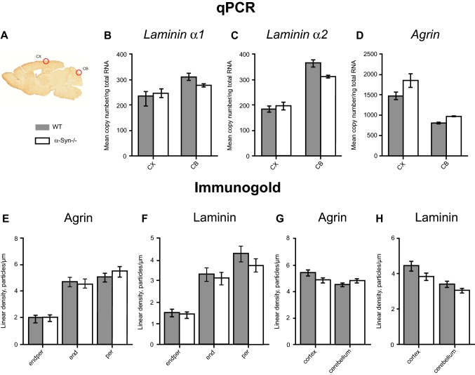
Evidence suggests that extracellular matrix molecules of perivascular basal laminae help orchestrate the molecular assemblies at the gliovascular interface. Specifically, laminin and agrin are thought to tether the dystrophin-associated protein (DAP) complex to the astrocytic basal lamina. This complex includes α-syntrophin (α-Syn), which is believed to anchor aquaporin-4 (AQP4) to astrocytic endfoot membrane domains. We have previously shown that the size of the perivascular AQP4 pool differs considerably between brain regions in an α-Syn-dependent manner. Also, both AQP4 and α-Syn occur at higher densities in endfoot membrane domains facing pericytes than in endfoot membrane domains facing endothelial cells. The heterogeneous distribution of AQP4 at the regional and capillary level has been attributed to a direct interaction between AQP4 and α-Syn. This would be challenged (1) if the microdistributions of laminin and agrin fail to align with those of DAP and AQP4 and (2) if targeted deletion of α-Syn leads to a loss of laminin and/or agrin. Here, we provide the first detailed and quantitative analysis of laminin and agrin in brain basal laminae of mice. We show that the microdistributions of these molecules vary in a fashion that is well aligned with the previously reported microdistribution of AQP4. We also demonstrate that the expression patterns of laminin and agrin are insensitive to targeted deletion of α-Syn, suggesting that α-Syn deletion affects AQP4 directly and not indirectly via laminin or agrin. These data fill remaining voids in the current model of how key molecules are assembled and tethered at the gliovascular interface.
有证据表明,血管周围基底层的细胞外基质分子有助于协调神经胶质血管界面的分子组装。具体来说,层粘连蛋白和聚集素被认为将营养不良相关蛋白 (DAP) 复合物固定在星形胶质细胞基底层上。该复合物包括α-突触核蛋白 (α-Syn),它被认为将水通道蛋白-4 (AQP4) 锚定在星形胶质细胞足突膜域。我们之前已经表明,AQP4 在血管周围的池大小在不同脑区之间以α-Syn 依赖的方式有很大差异。此外,AQP4 和 α-Syn 在面向周细胞的足突膜域中的密度都高于面向内皮细胞的足突膜域。AQP4 在区域和毛细血管水平的异质分布归因于 AQP4 和 α-Syn 之间的直接相互作用。如果层粘连蛋白和聚集素的微分布不能与 DAP 和 AQP4 的微分布对齐(1),或者如果靶向敲除 α-Syn 导致层粘连蛋白和/或聚集素丢失(2),这将受到挑战。在这里,我们提供了关于小鼠脑基底膜中层粘连蛋白和聚集素的首次详细和定量分析。我们表明,这些分子的微分布以与先前报道的 AQP4 微分布非常一致的方式变化。我们还证明,层粘连蛋白和聚集素的表达模式对靶向敲除 α-Syn 不敏感,这表明 α-Syn 缺失直接影响 AQP4,而不是通过层粘连蛋白或聚集素间接影响 AQP4。这些数据填补了当前神经胶质血管界面关键分子组装和固定模型中的剩余空白。