Department of Gynecology and Obstetrics, Botucatu Medical School (FMB), São Paulo State University (UNESP), CEP18618-687, Sao Paulo, Brazil.
Physics Department, Institute of Biosciences, Letters and Exact Sciences, Multiuser Center for Biomolecular Innovation, UNESP-São Paulo State University, Sao Paulo, Brazil.
BMC Pregnancy Childbirth. 2020 Feb 19;20(1):117. doi: 10.1186/s12884-020-2749-x.
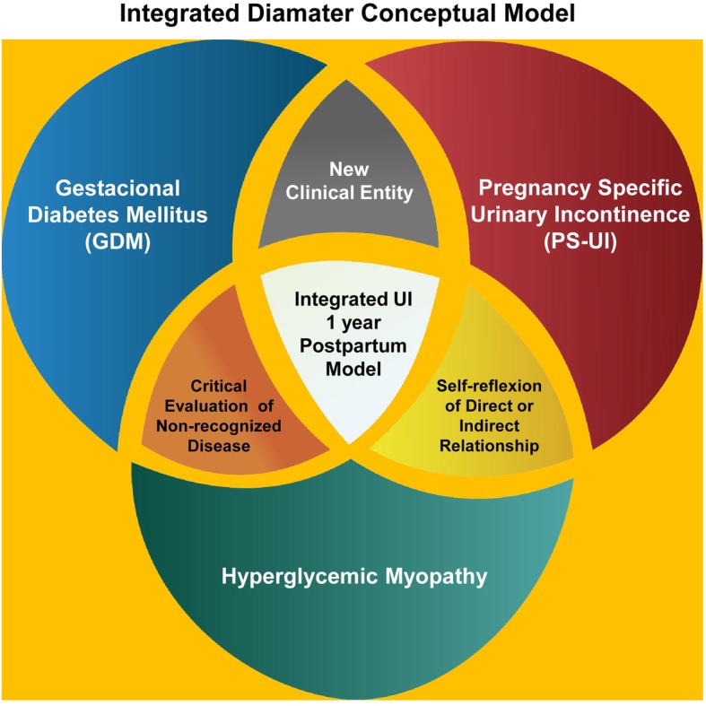
Pelvic floor muscles (PFM) and rectus abdominis muscles (RAM) of pregnant diabetic rats exhibit atrophy, co-localization of fast and slow fibers and an increased collagen type I/III ratio. However, the role of similar PFM or RAM hyperglycemic-related myopathy in women with gestational diabetes mellitus (GDM) remains poorly investigated. This study aims to assess the frequency of pelvic floor muscle disorders and pregnancy-specific urinary incontinence (PS-UI) 12 months after the Cesarean (C) section in women with GDM. Specifically, differences in PFM/RAM hyperglycemic myopathy will be evaluated.
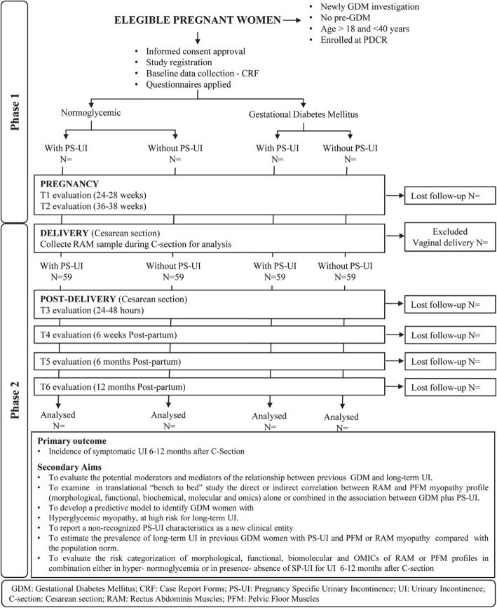
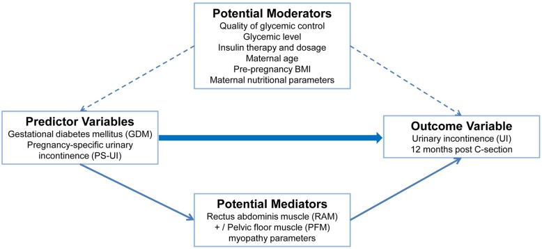
The Diamater is an ongoing cohort study of four groups of 59 pregnant women each from the Perinatal Diabetes Research Centre (PDRC), Botucatu Medical School (FMB)-UNESP (São Paulo State University), Brazil. Diagnosis of GDM and PS-UI will be made at 24-26 weeks, with a follow-up at 34-38 weeks of gestation. Inclusion in the study will occur at the time of C-section, and patients will be followed at 24-48 h, 6 weeks and 6 and 12 months postpartum. Study groups will be classified as (1) GDM plus PS-UI; (2) GDM without PS-UI; (3) Non-GDM plus PS-UI; and (4) Non-GDM without PS-UI. We will analyze relationships between GDM, PS-UI and hyperglycemic myopathy at 12 months after C-section. The mediator variables to be evaluated include digital palpation, vaginal squeeze pressure, 3D pelvic floor ultrasound, and 3D RAM ultrasound. RAM samples obtained during C-section will be analyzed for ex-vivo contractility, morphological, molecular and OMICS profiles to further characterize the hyperglycemic myopathy. Additional variables to be evaluated include maternal age, socioeconomic status, educational level, ethnicity, body mass index, weight gain during pregnancy, quality of glycemic control and insulin therapy.
To our knowledge, this will be the first study to provide data on the prevalence of PS-UI and RAM and PFM physical and biomolecular muscle profiles after C-section in mothers with GDM. The longitudinal design allows for the assessment of cause-effect relationships between GDM, PS-UI, and PFMs and RAMs myopathy. The findings may reveal previously undetermined consequences of GDM.
患有妊娠糖尿病的大鼠的盆底肌(PFM)和腹直肌(RAM)会出现萎缩、快肌和慢肌的共定位以及Ⅰ型/Ⅲ型胶原比例增加。然而,患有妊娠期糖尿病(GDM)的女性的类似 PFM 或 RAM 高血糖相关肌病的作用仍未得到充分研究。本研究旨在评估 GDM 产妇剖宫产(C)术后 12 个月时盆底肌功能障碍和妊娠特异性尿失禁(PS-UI)的发生频率。具体而言,将评估 PFM/RAM 高血糖性肌病的差异。
巴西博图卡图医学院(FMB)-南里奥格兰德州联邦大学(UNESP)围产期糖尿病研究中心(PDRC)正在进行一项前瞻性队列研究,共纳入四组各 59 名孕妇。GDM 和 PS-UI 的诊断将在 24-26 周进行,在妊娠 34-38 周进行随访。研究将在 C 剖宫产后纳入患者,患者将在 24-48 小时、6 周和 6 个月及 12 个月产后进行随访。研究组将分为(1)GDM 伴 PS-UI;(2)GDM 不伴 PS-UI;(3)非 GDM 伴 PS-UI;(4)非 GDM 不伴 PS-UI。我们将分析 C 剖宫产后 12 个月时 GDM、PS-UI 和高血糖性肌病之间的关系。将评估的中介变量包括数字触诊、阴道挤压压、3D 盆底超声和 3D RAM 超声。C 剖宫产后获得的 RAM 样本将进行离体收缩性、形态学、分子和 OMICS 分析,以进一步表征高血糖性肌病。还将评估其他变量,包括产妇年龄、社会经济地位、教育水平、种族、体重指数、孕期体重增加、血糖控制质量和胰岛素治疗。
据我们所知,这将是第一项研究,提供关于 GDM 产妇 C 剖宫产后 PS-UI 和 RAM 以及 PFM 物理和生物分子肌肉特征的患病率数据。纵向设计允许评估 GDM、PS-UI 和 PFM 与 RAM 肌病之间的因果关系。研究结果可能揭示 GDM 之前未确定的后果。