Cansu Aysegul, Bekircavusoglu Suleyman, Oguz Sukru, Bulut Eser, Fidan Sami
Karadeniz Technical University, Faculty of Medicine, Department of Radiology.
Trabzon Kanuni Education and Research Hospital, Department of Radiology.
Medicine (Baltimore). 2020 Feb;99(8):e19202. doi: 10.1097/MD.0000000000019202.
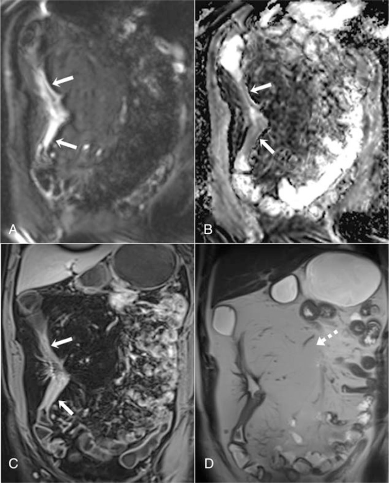
The present study aimed to investigate the potential use of T2-weighted sequences with diffusion weighted imaging (DWI) in magnetic resonance (MR) enterography instead of conventional contrast-enhanced MR imaging (MRI) sequences for the evaluation of active inflammation in Crohn disease.Two-hundred thirteen intestinal segments of 43 patients, who underwent colonoscopy within 2 weeks before or after MR enterography were evaluated in this retrospective study. DWI sequences, T2-weighted sequences, and contrast-enhanced T1-weighted sequences were acquired in the MR enterography scan after cleaning of the bowel and using an oral contrast agent. First, the intestinal segments that had active inflammation in MR enterography were qualitatively evaluated in T2-weighted and contrast-enhanced T1-weighted sequences and then MR activity index (MRAI 1) and MRAI 2 were formed with and without contrast-enhanced sequences in 2 separate sessions.The correlation coefficient between contrast enhanced and DWI MR enterography scores (MRAI 1 and MRAI 2) of intestinal inflammation was 0.97 for all segments. In addition, separate correlation coefficients were calculated for terminal ileum, right colon, transverse colon, left colon, and rectum, and there was a strong correlation between the MRAI 1 and MRAI 2 scores of each segment (r = 0.86-0.97, P < .001). On the other hand, MR enterography had 88.7% sensitivity, 97.9% specificity, 95.5% positive predictive value, 94.6% negative predictive value, and 94.8% accuracy for detection of active inflammation in all intestinal segments in Crohn disease.DWI and T2-weighted sequences acquired with cleaning of the bowel can be used instead of contrast-enhanced MRI sequences for the evaluation of active inflammation in Crohn disease.
本研究旨在探讨磁共振(MR)小肠造影中使用T2加权序列联合扩散加权成像(DWI)而非传统的对比增强MR成像(MRI)序列来评估克罗恩病的活动性炎症。在这项回顾性研究中,对43例在MR小肠造影前或后2周内接受结肠镜检查的患者的213个肠段进行了评估。在肠道清洁并使用口服对比剂后,于MR小肠造影扫描中采集DWI序列、T2加权序列和对比增强T1加权序列。首先,在T2加权和对比增强T1加权序列中对MR小肠造影中有活动性炎症的肠段进行定性评估,然后在两个独立的环节中分别使用和不使用对比增强序列形成MR活动指数(MRAI 1)和MRAI 2。所有肠段的肠道炎症对比增强和DWI MR小肠造影评分(MRAI 1和MRAI 2)之间的相关系数为0.97。此外,还分别计算了回肠末端、右半结肠、横结肠、左半结肠和直肠的相关系数,各段的MRAI 1和MRAI 2评分之间存在强相关性(r = 0.86 - 0.97,P <.001)。另一方面,MR小肠造影在检测克罗恩病所有肠段的活动性炎症方面具有88.7%的敏感性、97.9%的特异性、95.5%的阳性预测值、94.6%的阴性预测值和94.8%的准确性。肠道清洁后采集的DWI和T2加权序列可用于替代对比增强MRI序列来评估克罗恩病的活动性炎症。