• 文献检索
  • 文档翻译
  • 深度研究
  • 学术资讯
  • Suppr Zotero 插件Zotero 插件
  • 邀请有礼
  • 套餐&价格
  • 历史记录
应用&插件
Suppr Zotero 插件Zotero 插件浏览器插件Mac 客户端Windows 客户端微信小程序
定价
高级版会员购买积分包购买API积分包
服务
文献检索文档翻译深度研究API 文档MCP 服务
关于我们
关于 Suppr公司介绍联系我们用户协议隐私条款
关注我们

Suppr 超能文献

核心技术专利:CN118964589B侵权必究
粤ICP备2023148730 号-1Suppr @ 2026

文献检索

告别复杂PubMed语法,用中文像聊天一样搜索,搜遍4000万医学文献。AI智能推荐,让科研检索更轻松。

立即免费搜索

文件翻译

保留排版,准确专业,支持PDF/Word/PPT等文件格式,支持 12+语言互译。

免费翻译文档

深度研究

AI帮你快速写综述,25分钟生成高质量综述,智能提取关键信息,辅助科研写作。

立即免费体验

表观扩散系数测量在鉴别腋窝良恶性淋巴结中的应用

Measurement of apparent diffusion coefficient in discrimination of benign and malignant axillary lymph nodes.

作者信息

Yılmaz Ebru, Erok Berrin, Atca Ali Önder

机构信息

Sarıyer Hamidiye Etfal Education and Research Hospital, İstanbul, Turkey.

Cihanbeyli State Hospital, Konya, Turkey.

出版信息

Pol J Radiol. 2019 Dec 22;84:e592-e597. doi: 10.5114/pjr.2019.92315. eCollection 2019.

DOI:10.5114/pjr.2019.92315
PMID:32082458
原文链接:https://pmc.ncbi.nlm.nih.gov/articles/PMC7016376/
Abstract

PURPOSE

We aimed to determine the contribution of the apparent diffusion coefficient (ADC) value in the detection of axillary lymph node metastasis.

MATERIAL AND METHODS

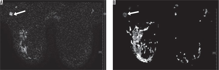
Breast magnetic resonance of 58 patients, performed in the radiology clinic of our hospital between 2015 and 2017 were examined retrospectively, and 43 lymph nodes in 43 patients were included in the study. They were evaluated morphologically on T1W and T2W sequences, and the lymph nodes showing rounded shape, focal or diffuse cortical thickness of more than 3 mm, and partial or total effacement of fatty hilum were included in the study. Subsequently, their ADC values were measured.

RESULTS

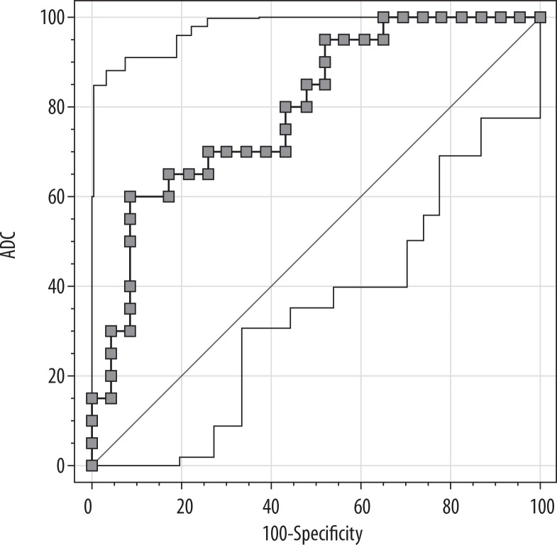
There were 43 lymph nodes, 20 of which were malignant and 23 of which were benign. While the mean ADC value of malignant axillary lymph nodes was 0.749 10 mm/s (0.48-1.342), it was 0.982 10 mm/s (0.552-1.986) for benign lymph nodes. When the ADC cut-off value was taken as ≤ 0.753 × 10 mm/s, its discrimination power between benign and malignant axillary lymph nodes was as follows: sensitivity - 60%; specificity - 91.3%; accuracy - 76.7%; positive predictive value - 85.7%; and negative predictive value - 72.4%.

CONCLUSIONS

There was no significant difference between mean ADC value of 12 lymphadenopathies (LAP) associated with inflammatory breast diseases (granulomatous mastitis and acute suppurative mastitis) and mean ADC value of metastatic lymph nodes. However, the ADC value of lymph nodes showing thickened cortex due to systemic inflammatory diseases was over 1, and there was a statistically significant difference when compared with metastatic lymph nodes.

摘要

目的

我们旨在确定表观扩散系数(ADC)值在腋窝淋巴结转移检测中的作用。

材料与方法

回顾性分析2015年至2017年在我院放射科进行的58例患者的乳腺磁共振成像,43例患者中的43个淋巴结纳入研究。在T1W和T2W序列上对其进行形态学评估,研究纳入形状呈圆形、局灶性或弥漫性皮质厚度超过3mm、脂肪门部分或完全消失的淋巴结。随后测量其ADC值。

结果

共有43个淋巴结,其中20个为恶性,23个为良性。恶性腋窝淋巴结的平均ADC值为0.749×10⁻³mm²/s(0.48 - 1.342),良性淋巴结的平均ADC值为0.982×10⁻³mm²/s(0.552 - 1.986)。当ADC截断值取为≤0.753×10⁻³mm²/s时,其区分良性和恶性腋窝淋巴结的能力如下:敏感性 - 60%;特异性 - 91.3%;准确性 - 76.7%;阳性预测值 - 85.7%;阴性预测值 - 72.4%。

结论

与炎性乳腺癌(肉芽肿性乳腺炎和急性化脓性乳腺炎)相关的12个淋巴结病变(LAP)的平均ADC值与转移性淋巴结的平均ADC值之间无显著差异。然而,因全身性炎症疾病导致皮质增厚的淋巴结的ADC值超过1,与转移性淋巴结相比有统计学显著差异。

https://cdn.ncbi.nlm.nih.gov/pmc/blobs/05cb/7016376/7f2efe097ce2/PJR-84-39575-g006.jpg
https://cdn.ncbi.nlm.nih.gov/pmc/blobs/05cb/7016376/0707b3446403/PJR-84-39575-g001.jpg
https://cdn.ncbi.nlm.nih.gov/pmc/blobs/05cb/7016376/52adebf9c1d7/PJR-84-39575-g002.jpg
https://cdn.ncbi.nlm.nih.gov/pmc/blobs/05cb/7016376/383197de5bf3/PJR-84-39575-g003.jpg
https://cdn.ncbi.nlm.nih.gov/pmc/blobs/05cb/7016376/a5351014f9c6/PJR-84-39575-g004.jpg
https://cdn.ncbi.nlm.nih.gov/pmc/blobs/05cb/7016376/c7ee02a15011/PJR-84-39575-g005.jpg
https://cdn.ncbi.nlm.nih.gov/pmc/blobs/05cb/7016376/7f2efe097ce2/PJR-84-39575-g006.jpg

相似文献

1
Measurement of apparent diffusion coefficient in discrimination of benign and malignant axillary lymph nodes.表观扩散系数测量在鉴别腋窝良恶性淋巴结中的应用
Pol J Radiol. 2019 Dec 22;84:e592-e597. doi: 10.5114/pjr.2019.92315. eCollection 2019.
2
Role of diffusion-weighted MRI: predicting axillary lymph node metastases in breast cancer.扩散加权磁共振成像的作用:预测乳腺癌腋窝淋巴结转移
Acta Radiol. 2014 Oct;55(8):909-16. doi: 10.1177/0284185113509094. Epub 2013 Nov 14.
3
Differentiation Between Benign and Metastatic Breast Lymph Nodes Using Apparent Diffusion Coefficients.利用表观扩散系数鉴别乳腺良性和转移性淋巴结
Front Oncol. 2022 Feb 23;12:795265. doi: 10.3389/fonc.2022.795265. eCollection 2022.
4
Diagnostic value of diffusion-weighted magnetic resonance imaging: differentiation of benign and malignant lymph nodes in different regions of the body.磁共振扩散加权成像的诊断价值:身体不同区域良恶性淋巴结的鉴别
Clin Imaging. 2015 Sep-Oct;39(5):856-62. doi: 10.1016/j.clinimag.2015.05.006. Epub 2015 May 22.
5
Apparent diffusion coefficient ratio between axillary lymph node with primary tumor to detect nodal metastasis in breast cancer patients.腋窝淋巴结与原发肿瘤的表观扩散系数比值在乳腺癌患者淋巴结转移检测中的应用。
J Magn Reson Imaging. 2013 Oct;38(4):824-8. doi: 10.1002/jmri.24031. Epub 2013 Feb 25.
6
Diagnostic Value of Diffusion Weighted Magnetic Resonance Imaging in Evaluation of Metastatic Axillary Lymph Nodes in a Sample of Iranian Women with Breast Cancer.扩散加权磁共振成像在评估伊朗乳腺癌女性样本中腋窝转移性淋巴结的诊断价值
Asian Pac J Cancer Prev. 2017 May 1;18(5):1265-1270. doi: 10.22034/APJCP.2017.18.5.1265.
7
Assessment of axillary lymph nodes in patients with breast cancer with diffusion-weighted MR imaging in combination with routine and dynamic contrast MR imaging.采用扩散加权磁共振成像联合常规及动态对比增强磁共振成像评估乳腺癌患者腋窝淋巴结。
Breast Cancer. 2016 May;23(3):525-32. doi: 10.1007/s12282-015-0598-7. Epub 2015 Mar 13.
8
Evaluation of the diagnostic performance of apparent diffusion coefficient (ADC) values on diffusion-weighted magnetic resonance imaging (DWI) in differentiating between benign and metastatic lymph nodes in cases of cholangiocarcinoma.评估磁共振扩散加权成像(DWI)表观扩散系数(ADC)值在胆管癌良恶性淋巴结鉴别诊断中的诊断性能。
Abdom Radiol (NY). 2019 Feb;44(2):473-481. doi: 10.1007/s00261-018-1742-6.
9
Diffusion tensor imaging and diffusion-weighted imaging on axillary lymph node status in breast cancer patients.乳腺癌患者腋窝淋巴结状态的弥散张量成像和弥散加权成像。
Diagn Interv Radiol. 2022 Jul;28(4):329-336. doi: 10.5152/dir.2022.21460.
10
Does axillary lymph node size predict better metastatic involvement than apparent diffusion coefficient (ADC) value in women with newly diagnosed breast cancer?在新诊断的乳腺癌女性中,腋窝淋巴结大小比表观扩散系数(ADC)值更能预测转移累及情况吗?
Acta Radiol. 2020 Nov;61(11):1494-1504. doi: 10.1177/0284185120903449. Epub 2020 Feb 16.

引用本文的文献

1
Diagnostic value of synthetic diffusion-weighted imaging on breast magnetic resonance imaging assessment: comparison with conventional diffusion-weighted imaging.合成扩散加权成像在乳腺磁共振成像评估中的诊断价值:与常规扩散加权成像的比较。
Diagn Interv Radiol. 2024 Mar 6;30(2):91-98. doi: 10.4274/dir.2023.232466. Epub 2023 Oct 27.
2
Multiparametric Magnetic Resonance Imaging in Evaluation of Benign and Malignant Breast Masses with Pathological Correlation.多参数磁共振成像在评估乳腺良恶性肿块中的应用及其与病理的相关性
Cureus. 2022 Feb 17;14(2):e22348. doi: 10.7759/cureus.22348. eCollection 2022 Feb.
3
Apparent diffusion coefficient magnetic resonance imaging (ADC-MRI) in the axillary breast cancer lymph node metastasis detection: a narrative review.

本文引用的文献

1
Stromal fibrosis: imaging features with diagnostic contribution of diffusion-weighted MRI.间质纤维化:扩散加权磁共振成像的影像学特征及其诊断价值
Br J Radiol. 2018 May;91(1085):20170706. doi: 10.1259/bjr.20170706. Epub 2018 Feb 16.
2
Diagnostic Value of Diffusion Weighted Magnetic Resonance Imaging in Evaluation of Metastatic Axillary Lymph Nodes in a Sample of Iranian Women with Breast Cancer.扩散加权磁共振成像在评估伊朗乳腺癌女性样本中腋窝转移性淋巴结的诊断价值
Asian Pac J Cancer Prev. 2017 May 1;18(5):1265-1270. doi: 10.22034/APJCP.2017.18.5.1265.
3
Assessment of axillary lymph nodes in patients with breast cancer with diffusion-weighted MR imaging in combination with routine and dynamic contrast MR imaging.
表观扩散系数磁共振成像(ADC-MRI)在腋窝乳腺癌淋巴结转移检测中的应用:一项叙述性综述。
Gland Surg. 2020 Dec;9(6):2225-2234. doi: 10.21037/gs-20-546.
采用扩散加权磁共振成像联合常规及动态对比增强磁共振成像评估乳腺癌患者腋窝淋巴结。
Breast Cancer. 2016 May;23(3):525-32. doi: 10.1007/s12282-015-0598-7. Epub 2015 Mar 13.
4
Diagnostic value of breast MRI for predicting metastatic axillary lymph nodes in breast cancer patients: diffusion-weighted MRI and conventional MRI.乳腺MRI对预测乳腺癌患者腋窝淋巴结转移的诊断价值:扩散加权MRI与传统MRI
Magn Reson Imaging. 2014 Dec;32(10):1230-6. doi: 10.1016/j.mri.2014.07.001. Epub 2014 Jul 26.
5
The value of T2* in differentiating metastatic from benign axillary lymph nodes in patients with breast cancer--a preliminary in vivo study.在乳腺癌患者中,T2* 值在鉴别转移性与良性腋窝淋巴结中的价值——一项初步的体内研究。
PLoS One. 2014 Jan 16;9(1):e84038. doi: 10.1371/journal.pone.0084038. eCollection 2014.
6
Role of diffusion-weighted MRI: predicting axillary lymph node metastases in breast cancer.扩散加权磁共振成像的作用:预测乳腺癌腋窝淋巴结转移
Acta Radiol. 2014 Oct;55(8):909-16. doi: 10.1177/0284185113509094. Epub 2013 Nov 14.
7
The role of apparent diffusion coefficient values in the differential diagnosis of breast lesions in diffusion-weighted MRI.表观扩散系数值在弥散加权 MRI 鉴别诊断乳腺病变中的作用。
Diagn Interv Radiol. 2013 Nov-Dec;19(6):457-62. doi: 10.5152/dir.2013.12132.
8
Axillary lymph node characterization in breast cancer patients using magnetic resonance mammography: a prospective comparative study with FDG PET-CT and healthy women.应用磁共振乳腺成像对乳腺癌患者腋窝淋巴结进行特征描述:与 FDG PET-CT 及健康女性对照的前瞻性研究。
Eur J Radiol. 2013 Dec;82(12):2194-8. doi: 10.1016/j.ejrad.2013.08.010. Epub 2013 Aug 12.
9
Can additional histopathological examination of ultrasound-guided axillary lymph node core biopsies improve preoperative diagnosis of primary breast cancer nodal metastasis?超声引导下腋窝淋巴结核心活检的额外组织病理学检查能否改善原发性乳腺癌淋巴结转移的术前诊断?
Clin Radiol. 2013 Jul;68(7):704-7. doi: 10.1016/j.crad.2013.01.020. Epub 2013 Mar 19.
10
Differential diagnosis of axillary inflammatory and metastatic lymph nodes in rabbit models by using diffusion-weighted imaging: compared with conventional magnetic resonance imaging.采用弥散加权成像对兔模型腋窝炎性及转移性淋巴结进行鉴别诊断:与常规磁共振成像比较。
Korean J Radiol. 2012 Jul-Aug;13(4):458-66. doi: 10.3348/kjr.2012.13.4.458. Epub 2012 Jun 18.