• 文献检索
  • 文档翻译
  • 深度研究
  • 学术资讯
  • Suppr Zotero 插件Zotero 插件
  • 邀请有礼
  • 套餐&价格
  • 历史记录
应用&插件
Suppr Zotero 插件Zotero 插件浏览器插件Mac 客户端Windows 客户端微信小程序
定价
高级版会员购买积分包购买API积分包
服务
文献检索文档翻译深度研究API 文档MCP 服务
关于我们
关于 Suppr公司介绍联系我们用户协议隐私条款
关注我们

Suppr 超能文献

核心技术专利:CN118964589B侵权必究
粤ICP备2023148730 号-1Suppr @ 2026

文献检索

告别复杂PubMed语法,用中文像聊天一样搜索,搜遍4000万医学文献。AI智能推荐,让科研检索更轻松。

立即免费搜索

文件翻译

保留排版,准确专业,支持PDF/Word/PPT等文件格式,支持 12+语言互译。

免费翻译文档

深度研究

AI帮你快速写综述,25分钟生成高质量综述,智能提取关键信息,辅助科研写作。

立即免费体验

扩散加权磁共振成像在评估伊朗乳腺癌女性样本中腋窝转移性淋巴结的诊断价值

Diagnostic Value of Diffusion Weighted Magnetic Resonance Imaging in Evaluation of Metastatic Axillary Lymph Nodes in a Sample of Iranian Women with Breast Cancer.

作者信息

Hasanzadeh Fereshteh, Faeghi Fariborz, Valizadeh Abdollah, Bayani Leyla

机构信息

Radiology Technology Department, School of Allied Medical Sciences, Shahid Beheshti University of Medical Sciences, Tehran, Iran. Email:

出版信息

Asian Pac J Cancer Prev. 2017 May 1;18(5):1265-1270. doi: 10.22034/APJCP.2017.18.5.1265.

DOI:10.22034/APJCP.2017.18.5.1265
PMID:28610412
原文链接:https://pmc.ncbi.nlm.nih.gov/articles/PMC5555533/
Abstract

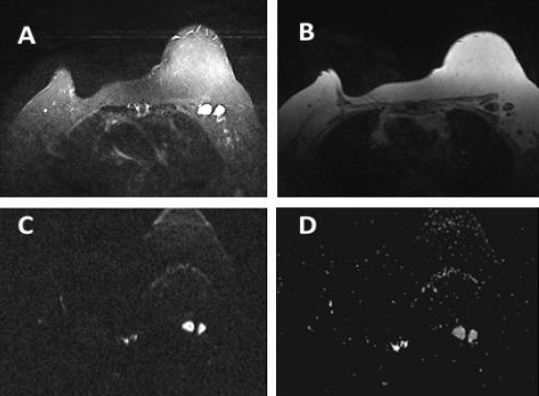
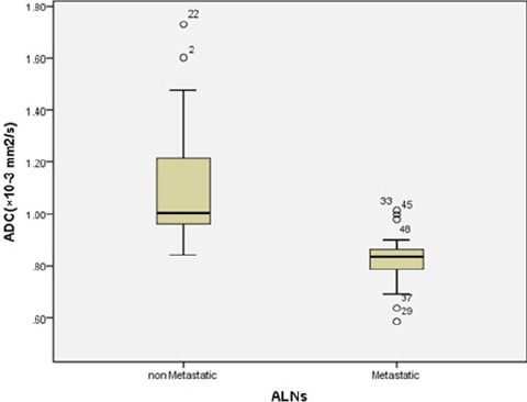
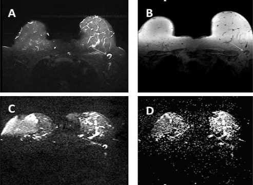
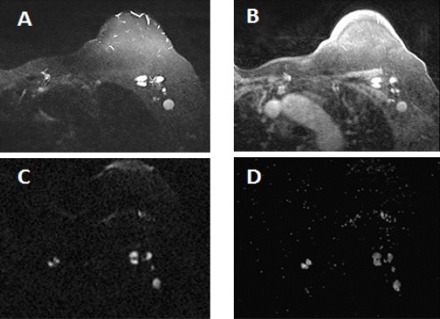
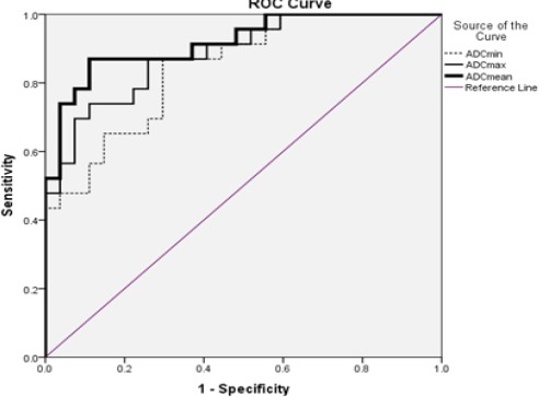
Objective: To evaluate the diagnostic value of diffusion weighted magnetic resonance imaging (DW-MRI) in assessment of metastases in axillary lymph nodes (ALNs) in a sample of Iranian women with breast cancer. Methods: A total of 50 axillary lymph nodes from 30 female patients with histologically verified breast cancer were assessed by 1.5 T MRI. DWI was implemented at b-values of 50, 400 and 800 s/mm2. Short axis diameter, presence of fatty hilum and apparent diffusion coefficient (ADC) values (min, max and mean) of metastatic and non-metastatic ALNs was compared. Cutoff ADC values to discriminate between benign and malignant axillary lymph nodes were analyzed with receiver coefficient characteristic (ROC) curves. Result: The final histopathological examination revealed 46% (n=23) metastatic and 54% (n=27) non-metastatic ALNs. There was no statistically significant difference in short axis diameter between the two groups (p = 0.537). However there was significantly correlation between loss of fatty hilum and presence of metastases (p < 0.001) and ADC values (0.255 ± 0.19×10⁻³ mm2/s vs 0.616 ±0.3×10⁻³ mm2/s (ADC min), 1.088 ± 0.22×10⁻³ mm2/s vs 1.497 ± 0.24×10⁻³ mm2/s (ADC max) and 0.824 ± 0.103 ×10⁻³ mm2/s vs 1.098 ± 0.23 ×10⁻³ mm2/s (ADC mean)) of metastatic ALNs were significantly lower than those of non-metastatic ALNs (p < 0.001). The optimal mean ADC cut-off value for differentiation between metastatic and non-metastatic ALNs was 0.904×10⁻³ mm2/s which had a higher specificity (88.9%) and accuracy (91.8%) as compared with ADC min and ADC max. Conclusion: DWI-MRI and ADC values are promising imaging methods which can assess metastatic ALNs in breast cancer with high sensitivity, specificity and accuracy.

摘要

目的

评估扩散加权磁共振成像(DW-MRI)在评估伊朗乳腺癌女性样本腋窝淋巴结(ALNs)转移中的诊断价值。方法:对30例经组织学证实为乳腺癌的女性患者的50个腋窝淋巴结进行1.5 T MRI评估。在b值为50、400和800 s/mm²时进行DWI。比较转移性和非转移性ALNs的短轴直径、脂肪门的存在情况以及表观扩散系数(ADC)值(最小值、最大值和平均值)。用受试者工作特征(ROC)曲线分析区分良性和恶性腋窝淋巴结的ADC临界值。结果:最终组织病理学检查显示46%(n = 23)为转移性ALNs,54%(n = 27)为非转移性ALNs。两组间短轴直径无统计学显著差异(p = 0.537)。然而,脂肪门缺失与转移的存在之间存在显著相关性(p < 0.001),转移性ALNs的ADC值(ADC最小值:0.255±0.19×10⁻³ mm²/s对0.616±0.3×10⁻³ mm²/s,ADC最大值:1.088±0.22×10⁻³ mm²/s对1.497±0.24×10⁻³ mm²/s,ADC平均值:0.824±0.103×10⁻³ mm²/s对1.098±0.23×10⁻³ mm²/s)显著低于非转移性ALNs(p < 0.001)。转移性和非转移性ALNs鉴别的最佳平均ADC临界值为0.904×10⁻³ mm²/s,与ADC最小值和ADC最大值相比,其具有更高的特异性(88.9%)和准确性(91.8%)。结论:DWI-MRI和ADC值是有前景的成像方法,可高灵敏度、特异性和准确性地评估乳腺癌中的转移性ALNs。

https://cdn.ncbi.nlm.nih.gov/pmc/blobs/aa8c/5555533/9469a0e1db7f/APJCP-18-1265-g005.jpg
https://cdn.ncbi.nlm.nih.gov/pmc/blobs/aa8c/5555533/fdeaaf5093bb/APJCP-18-1265-g001.jpg
https://cdn.ncbi.nlm.nih.gov/pmc/blobs/aa8c/5555533/3407411981ea/APJCP-18-1265-g002.jpg
https://cdn.ncbi.nlm.nih.gov/pmc/blobs/aa8c/5555533/12ca51e6051e/APJCP-18-1265-g003.jpg
https://cdn.ncbi.nlm.nih.gov/pmc/blobs/aa8c/5555533/f7cbbae0fd97/APJCP-18-1265-g004.jpg
https://cdn.ncbi.nlm.nih.gov/pmc/blobs/aa8c/5555533/9469a0e1db7f/APJCP-18-1265-g005.jpg

相似文献

1
Diagnostic Value of Diffusion Weighted Magnetic Resonance Imaging in Evaluation of Metastatic Axillary Lymph Nodes in a Sample of Iranian Women with Breast Cancer.扩散加权磁共振成像在评估伊朗乳腺癌女性样本中腋窝转移性淋巴结的诊断价值
Asian Pac J Cancer Prev. 2017 May 1;18(5):1265-1270. doi: 10.22034/APJCP.2017.18.5.1265.
2
Diagnostic Value of Diffusion Weighted Magnetic Resonance Imaging in Evaluation of Metastatic Neck Lymph Nodes in Head and Neck Cancer: A Sample of Iranian Patient.扩散加权磁共振成像在评估头颈癌颈部转移性淋巴结中的诊断价值:以伊朗患者为例
Asian Pac J Cancer Prev. 2019 Jun 1;20(6):1789-1795. doi: 10.31557/APJCP.2019.20.6.1789.
3
Role of diffusion-weighted MRI: predicting axillary lymph node metastases in breast cancer.扩散加权磁共振成像的作用:预测乳腺癌腋窝淋巴结转移
Acta Radiol. 2014 Oct;55(8):909-16. doi: 10.1177/0284185113509094. Epub 2013 Nov 14.
4
Diffusion tensor imaging and diffusion-weighted imaging on axillary lymph node status in breast cancer patients.乳腺癌患者腋窝淋巴结状态的弥散张量成像和弥散加权成像。
Diagn Interv Radiol. 2022 Jul;28(4):329-336. doi: 10.5152/dir.2022.21460.
5
Diagnostic Usefulness of Diffusion-Weighted MRI for Axillary Lymph Node Evaluation in Patients with Breast Cancer.扩散加权磁共振成像在乳腺癌患者腋窝淋巴结评估中的诊断价值
Diagnostics (Basel). 2023 Jan 31;13(3):513. doi: 10.3390/diagnostics13030513.
6
Apparent diffusion coefficient ratio between axillary lymph node with primary tumor to detect nodal metastasis in breast cancer patients.腋窝淋巴结与原发肿瘤的表观扩散系数比值在乳腺癌患者淋巴结转移检测中的应用。
J Magn Reson Imaging. 2013 Oct;38(4):824-8. doi: 10.1002/jmri.24031. Epub 2013 Feb 25.
7
[Diagnosis value of 3.0 T diffusion-weighted imaging with background suppression magnetic resonance for metastatic lymph nodes in rectal cancer].3.0T背景抑制磁共振扩散加权成像对直肠癌转移性淋巴结的诊断价值
Zhonghua Wei Chang Wai Ke Za Zhi. 2011 Nov;14(11):842-5.
8
Assessment of axillary lymph nodes in patients with breast cancer with diffusion-weighted MR imaging in combination with routine and dynamic contrast MR imaging.采用扩散加权磁共振成像联合常规及动态对比增强磁共振成像评估乳腺癌患者腋窝淋巴结。
Breast Cancer. 2016 May;23(3):525-32. doi: 10.1007/s12282-015-0598-7. Epub 2015 Mar 13.
9
Evaluation of the diagnostic performance of apparent diffusion coefficient (ADC) values on diffusion-weighted magnetic resonance imaging (DWI) in differentiating between benign and metastatic lymph nodes in cases of cholangiocarcinoma.评估磁共振扩散加权成像(DWI)表观扩散系数(ADC)值在胆管癌良恶性淋巴结鉴别诊断中的诊断性能。
Abdom Radiol (NY). 2019 Feb;44(2):473-481. doi: 10.1007/s00261-018-1742-6.
10
Diffusion weighted images of metastatic as compared with nonmetastatic axillary lymph nodes in patients with newly diagnosed breast cancer.新诊断乳腺癌患者中转移性腋窝淋巴结与非转移性腋窝淋巴结的扩散加权成像。
J Magn Reson Imaging. 2015 Sep;42(3):771-8. doi: 10.1002/jmri.24829. Epub 2014 Dec 30.

引用本文的文献

1
Prediction of axillary nodal burden using preoperative magnetic resonance imaging scoring in patients with clinically node-negative breast cancer: a retrospective cohort study.术前磁共振成像评分预测临床腋窝淋巴结阴性乳腺癌患者的腋窝淋巴结负荷:一项回顾性队列研究。
Gland Surg. 2024 Dec 31;13(12):2288-2299. doi: 10.21037/gs-24-379. Epub 2024 Dec 27.
2
Differentiation Between Benign and Metastatic Breast Lymph Nodes Using Apparent Diffusion Coefficients.利用表观扩散系数鉴别乳腺良性和转移性淋巴结
Front Oncol. 2022 Feb 23;12:795265. doi: 10.3389/fonc.2022.795265. eCollection 2022.
3
Measurement of apparent diffusion coefficient in discrimination of benign and malignant axillary lymph nodes.

本文引用的文献

1
Comparative Investigation of Single Voxel Magnetic Resonance Spectroscopy and Dynamic Contrast Enhancement MR Imaging in Differentiation of Benign and Malignant Breast Lesions in a Sample of Iranian Women.单体素磁共振波谱与动态对比增强磁共振成像在伊朗女性样本中鉴别乳腺良恶性病变的对比研究
Asian Pac J Cancer Prev. 2015;16(18):8335-8. doi: 10.7314/apjcp.2015.16.18.8335.
2
Assessment of axillary lymph nodes in patients with breast cancer with diffusion-weighted MR imaging in combination with routine and dynamic contrast MR imaging.采用扩散加权磁共振成像联合常规及动态对比增强磁共振成像评估乳腺癌患者腋窝淋巴结。
Breast Cancer. 2016 May;23(3):525-32. doi: 10.1007/s12282-015-0598-7. Epub 2015 Mar 13.
3
表观扩散系数测量在鉴别腋窝良恶性淋巴结中的应用
Pol J Radiol. 2019 Dec 22;84:e592-e597. doi: 10.5114/pjr.2019.92315. eCollection 2019.
4
Diagnostic Value of Diffusion Weighted Magnetic Resonance Imaging in Evaluation of Metastatic Neck Lymph Nodes in Head and Neck Cancer: A Sample of Iranian Patient.扩散加权磁共振成像在评估头颈癌颈部转移性淋巴结中的诊断价值:以伊朗患者为例
Asian Pac J Cancer Prev. 2019 Jun 1;20(6):1789-1795. doi: 10.31557/APJCP.2019.20.6.1789.
5
Is there any relationship between adc values of diffusion-weighted imaging and the histopathological prognostic factors of invasive ductal carcinoma?扩散加权成像的表观扩散系数值与浸润性导管癌的组织病理学预后因素之间是否存在关联?
Br J Radiol. 2018 Apr;91(1084):20170705. doi: 10.1259/bjr.20170705. Epub 2018 Jan 12.
Diffusion weighted images of metastatic as compared with nonmetastatic axillary lymph nodes in patients with newly diagnosed breast cancer.
新诊断乳腺癌患者中转移性腋窝淋巴结与非转移性腋窝淋巴结的扩散加权成像。
J Magn Reson Imaging. 2015 Sep;42(3):771-8. doi: 10.1002/jmri.24829. Epub 2014 Dec 30.
4
Differential diagnosis between metastatic and non-metastatic lymph nodes using DW-MRI: a meta-analysis of diagnostic accuracy studies.使用扩散加权磁共振成像鉴别转移性和非转移性淋巴结:诊断准确性研究的荟萃分析
J Cancer Res Clin Oncol. 2015 Jun;141(6):1119-30. doi: 10.1007/s00432-014-1895-9. Epub 2014 Dec 17.
5
Diagnostic Performance of Dedicated Axillary T2- and Diffusion-weighted MR Imaging for Nodal Staging in Breast Cancer.专用腋窝 T2 加权和扩散加权 MRI 对乳腺癌淋巴结分期的诊断性能。
Radiology. 2015 May;275(2):345-55. doi: 10.1148/radiol.14141167. Epub 2014 Dec 15.
6
Diagnostic value of breast MRI for predicting metastatic axillary lymph nodes in breast cancer patients: diffusion-weighted MRI and conventional MRI.乳腺MRI对预测乳腺癌患者腋窝淋巴结转移的诊断价值:扩散加权MRI与传统MRI
Magn Reson Imaging. 2014 Dec;32(10):1230-6. doi: 10.1016/j.mri.2014.07.001. Epub 2014 Jul 26.
7
Accuracy of individual descriptors and grading of nodal involvement by axillary ultrasound in patients of breast cancer.乳腺癌患者腋窝超声检查中单个描述符的准确性及腋窝淋巴结受累情况分级
Int J Breast Cancer. 2013;2013:930596. doi: 10.1155/2013/930596. Epub 2013 Dec 19.
8
Role of diffusion-weighted MRI: predicting axillary lymph node metastases in breast cancer.扩散加权磁共振成像的作用:预测乳腺癌腋窝淋巴结转移
Acta Radiol. 2014 Oct;55(8):909-16. doi: 10.1177/0284185113509094. Epub 2013 Nov 14.
9
Diagnostic performance of 18F-FDG PET/CT, ultrasonography and MRI. Detection of axillary lymph node metastasis in breast cancer patients.18F-FDG PET/CT、超声检查和MRI的诊断性能。乳腺癌患者腋窝淋巴结转移的检测。
Nuklearmedizin. 2014;53(3):89-94. doi: 10.3413/Nukmed-0605-13-06. Epub 2013 Nov 13.
10
Can additional histopathological examination of ultrasound-guided axillary lymph node core biopsies improve preoperative diagnosis of primary breast cancer nodal metastasis?超声引导下腋窝淋巴结核心活检的额外组织病理学检查能否改善原发性乳腺癌淋巴结转移的术前诊断?
Clin Radiol. 2013 Jul;68(7):704-7. doi: 10.1016/j.crad.2013.01.020. Epub 2013 Mar 19.