Fardanesh Reza, Thakur Sunitha B, Sevilimedu Varadan, Horvat Joao V, Gullo Roberto Lo, Reiner Jeffrey S, Eskreis-Winkler Sarah, Thakur Nikita, Pinker Katja
Department of Radiology, Memorial Sloan Kettering Cancer Center, New York, NY, United States.
Department of Medical Physics, Memorial Sloan Kettering Cancer Center, New York, NY, United States.
Front Oncol. 2022 Feb 23;12:795265. doi: 10.3389/fonc.2022.795265. eCollection 2022.
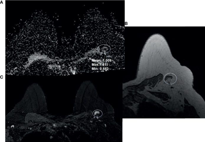
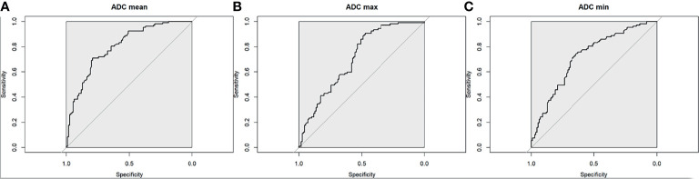
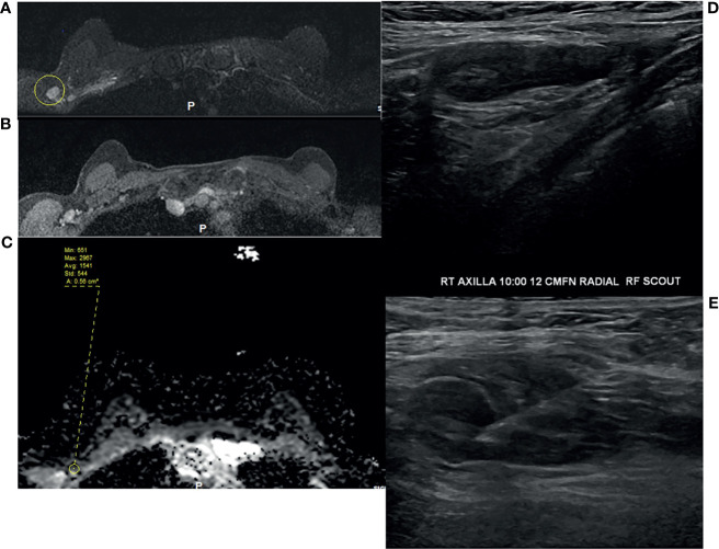
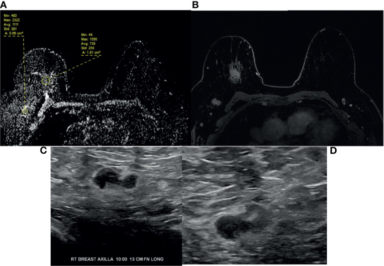
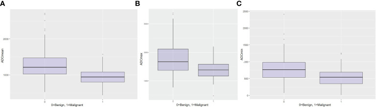
The aim of this study was to determine the range of apparent diffusion coefficient (ADC) values for benign axillary lymph nodes in contrast to malignant axillary lymph nodes, and to define the optimal ADC thresholds for three different ADC parameters (minimum, maximum, and mean ADC) in differentiating between benign and malignant lymph nodes. This retrospective study included consecutive patients who underwent breast MRI from January 2017-December 2020. Two-year follow-up breast imaging or histopathology served as the reference standard for axillary lymph node status. Area under the receiver operating characteristic curve (AUC) values for minimum, maximum, and mean ADC (min ADC, max ADC, and mean ADC) for benign malignant axillary lymph nodes were determined using the Wilcoxon rank sum test, and optimal ADC thresholds were determined using Youden's Index. The final study sample consisted of 217 patients (100% female, median age of 52 years (range, 22-81), 110 with benign axillary lymph nodes and 107 with malignant axillary lymph nodes. For benign axillary lymph nodes, ADC values (×10 mm/s) ranged from 0.522-2.712 for mean ADC, 0.774-3.382 for max ADC, and 0.071-2.409 for min ADC; for malignant axillary lymph nodes, ADC values (×10 mm/s) ranged from 0.796-1.080 for mean ADC, 1.168-1.592 for max ADC, and 0.351-0.688 for min ADC for malignant axillary lymph nodes. While there was a statistically difference in all ADC parameters (p<0.001) between benign and malignant axillary lymph nodes, boxplots illustrate overlaps in ADC values, with the least overlap occurring with mean ADC, suggesting that this is the most useful ADC parameter for differentiating between benign and malignant axillary lymph nodes. The mean ADC threshold that resulted in the highest diagnostic accuracy for differentiating between benign and malignant lymph nodes was 1.004×10 mm/s, yielding an accuracy of 75%, sensitivity of 71%, specificity of 79%, positive predictive value of 77%, and negative predictive value of 74%. This mean ADC threshold is lower than the European Society of Breast Imaging (EUSOBI) mean ADC threshold of 1.300×10 mm/s, therefore suggesting that the EUSOBI threshold which was recently recommended for breast tumors should not be extrapolated to evaluate the axillary lymph nodes.
本研究的目的是确定良性腋窝淋巴结与恶性腋窝淋巴结表观扩散系数(ADC)值的范围,并确定三种不同ADC参数(最小、最大和平均ADC)在区分良性和恶性淋巴结时的最佳ADC阈值。这项回顾性研究纳入了2017年1月至2020年12月期间连续接受乳腺MRI检查的患者。两年的随访乳腺成像或组织病理学检查作为腋窝淋巴结状态的参考标准。使用Wilcoxon秩和检验确定良性和恶性腋窝淋巴结的最小、最大和平均ADC(min ADC、max ADC和mean ADC)的受试者操作特征曲线下面积(AUC)值,并使用约登指数确定最佳ADC阈值。最终的研究样本包括217名患者(100%为女性,中位年龄52岁(范围22 - 81岁)),其中110例腋窝淋巴结为良性,107例腋窝淋巴结为恶性。对于良性腋窝淋巴结,平均ADC的ADC值(×10⁻³mm²/s)范围为0.522 - 2.712,最大ADC为0.774 - 3.382,最小ADC为0.071 - 2.409;对于恶性腋窝淋巴结,平均ADC的ADC值(×10⁻³mm²/s)范围为0.796 - 1.080,最大ADC为1.168 - 1.592,最小ADC为0.351 - 0.688。虽然良性和恶性腋窝淋巴结在所有ADC参数上均存在统计学差异(p<0.001),但箱线图显示ADC值存在重叠,平均ADC的重叠最少,这表明平均ADC是区分良性和恶性腋窝淋巴结最有用的ADC参数。区分良性和恶性淋巴结诊断准确性最高的平均ADC阈值为1.004×10⁻³mm²/s,准确率为75%,灵敏度为71%,特异性为79%,阳性预测值为77%,阴性预测值为74%。这个平均ADC阈值低于欧洲乳腺影像学会(EUSOBI)的平均ADC阈值1.300×10⁻³mm²/s,因此表明最近推荐用于乳腺肿瘤的EUSOBI阈值不应外推用于评估腋窝淋巴结。