Bede Peter, Chipika Rangariroyashe H, Finegan Eoin, Li Hi Shing Stacey, Chang Kai Ming, Doherty Mark A, Hengeveld Jennifer C, Vajda Alice, Hutchinson Siobhan, Donaghy Colette, McLaughlin Russell L, Hardiman Orla
Computational Neuroimaging Group, Biomedical Sciences Institute, Trinity College Dublin, 152-160 Pearse Street, Dublin 2, Ireland.
Electronics and Computer Science, University of Southampton, Southampton, SO17 1BJ, United Kingdom.
Data Brief. 2020 Feb 3;29:105229. doi: 10.1016/j.dib.2020.105229. eCollection 2020 Apr.
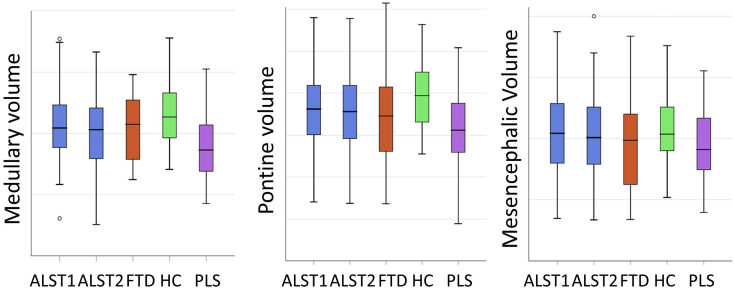
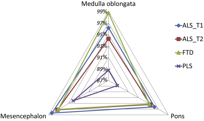
A standardised, single-centre, longitudinal imaging protocol was used to evaluate longitudinal brainstem alterations in 100 patients with amyotrophic lateral sclerosis (ALS) with reference to 33 patients with primary lateral sclerosis (PLS), 30 patients with frontotemporal dementia (FTD) and 100 healthy controls. "Brainstem pathology in amyotrophic lateral sclerosis and primary lateral sclerosis: A longitudinal neuroimaging study" [1] ALS patients were scanned twice; 4 months apart. T1-weighted imaging data were acquired on a 3 T Philips Achieva MRI system, using a 3D Inversion Recovery prepared Spoiled Gradient Recalled echo (IR-SPGR) sequence. Raw MRI data underwent meticulous quality control before pre-processing. A Bayesian segmentation algorithm was utilised to parcellate the brainstem into the medulla oblongata, pons and mesencephalon before estimating the volume of each segment. Vertex-based shape analyses were carried out to characterise anatomical patterns of atrophy. Brainstem volume loss in ALS was dominated by medulla oblongata atrophy, but significant pontine pathology was also detected. Brainstem volume reductions were more significant in PLS than in ALS after correcting for demographic variables and total intracranial volume. Shape analyses revealed bilateral 'flattening' of the medullary pyramids in ALS compared to healthy controls. Our data demonstrate that computational neuroimaging readily detects brainstem pathology in vivo in both amyotrophic lateral sclerosis and primary lateral sclerosis.
采用标准化的单中心纵向成像方案,以33例原发性侧索硬化症(PLS)患者、30例额颞叶痴呆(FTD)患者和100名健康对照为参照,评估100例肌萎缩侧索硬化症(ALS)患者脑干的纵向改变。“肌萎缩侧索硬化症和原发性侧索硬化症的脑干病理学:一项纵向神经影像学研究”[1]对ALS患者进行了两次扫描,间隔4个月。在3T飞利浦Achieva MRI系统上使用三维反转恢复预脉冲扰相梯度回波(IR-SPGR)序列采集T1加权成像数据。在预处理之前,对原始MRI数据进行了细致的质量控制。在估计每个节段的体积之前,利用贝叶斯分割算法将脑干分割为延髓、脑桥和中脑。进行基于顶点的形状分析以表征萎缩的解剖模式。ALS患者的脑干体积损失以延髓萎缩为主,但也检测到明显的脑桥病变。在校正人口统计学变量和总颅内体积后,PLS患者的脑干体积减少比ALS患者更显著。形状分析显示,与健康对照相比,ALS患者延髓锥体出现双侧“变平”。我们的数据表明,计算机神经成像能够在活体中轻松检测出肌萎缩侧索硬化症和原发性侧索硬化症患者的脑干病变。