Computational Neuroimaging Group (CNG), School of Medicine, Trinity College Dublin, Dublin 2, Ireland.
Department of Neurology, St James's Hospital, Dublin, Ireland.
J Neurol. 2024 Jun;271(6):3239-3255. doi: 10.1007/s00415-024-12261-z. Epub 2024 Mar 5.
Primary lateral sclerosis (PLS) is traditionally solely associated with progressive upper motor neuron dysfunction manifesting in limb spasticity, gait impairment, bulbar symptoms and pseudobulbar affect. Recent studies have described frontotemporal dysfunction in some patients resulting in cognitive manifestations. Cerebellar pathology is much less well characterised despite sporadic reports of cerebellar disease.
A multi-timepoint, longitudinal neuroimaging study was conducted to characterise the evolution of both intra-cerebellar disease burden and cerebro-cerebellar connectivity. The volumes of deep cerebellar nuclei, cerebellar cortical volumes, cerebro-cerebellar structural and functional connectivity were assessed longitudinally in a cohort of 43 individuals with PLS.
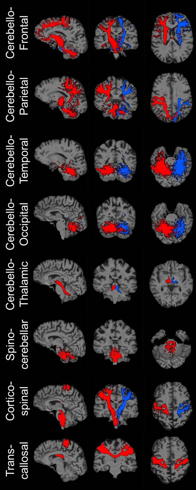
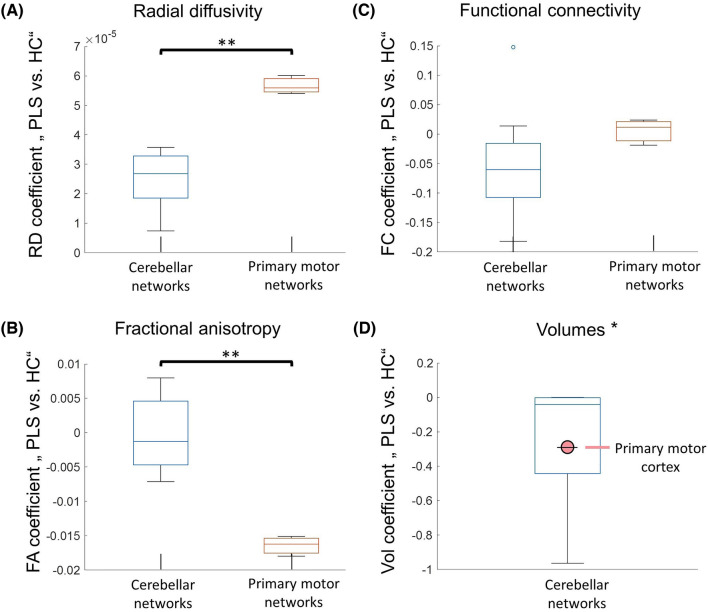
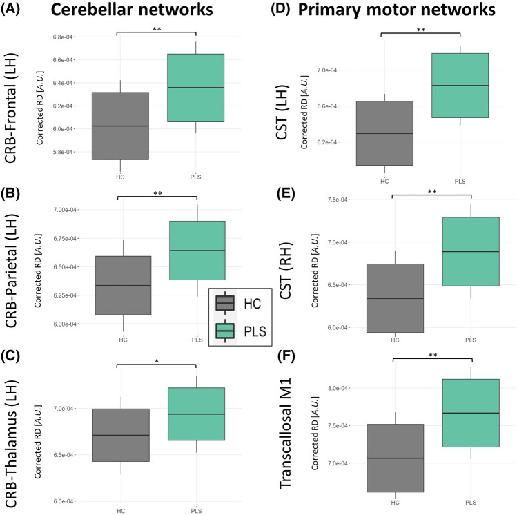
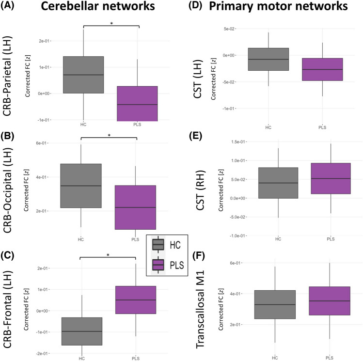
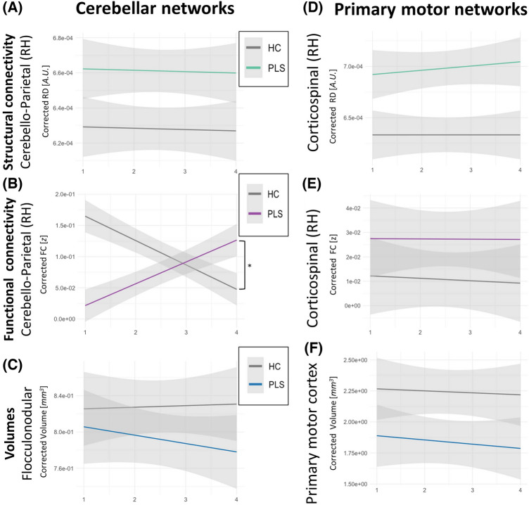
Cerebello-frontal, -temporal, -parietal, -occipital and cerebello-thalamic structural disconnection was detected at baseline based on radial diffusivity (RD) and cerebello-frontal decoupling was also evident based on fractional anisotropy (FA) alterations. Functional connectivity changes were also detected in cerebello-frontal, parietal and occipital projections. Volume reductions were identified in the vermis, anterior lobe, posterior lobe, and crura. Among the deep cerebellar nuclei, the dorsal dentate was atrophic. Longitudinal follow-up did not capture statistically significant progressive changes. Significant primary motor cortex atrophy and inter-hemispheric transcallosal degeneration were also captured.
PLS is not only associated with upper motor neuron dysfunction, but cerebellar cortical volume loss and deep cerebellar nuclear atrophy can also be readily detected. In addition to intra-cerebellar disease burden, cerebro-cerebellar connectivity alterations also take place. Our data add to the evolving evidence of widespread neurodegeneration in PLS beyond the primary motor regions. Cerebellar dysfunction in PLS is likely to exacerbate bulbar, gait and dexterity impairment and contribute to pseudobulbar affect.
原发性侧索硬化症(PLS)传统上仅与进行性上运动神经元功能障碍相关,表现为肢体痉挛、步态障碍、延髓症状和假性延髓影响。最近的研究描述了一些患者的额颞叶功能障碍,导致认知表现。尽管有零星的小脑疾病报告,但小脑病理特征描述得较少。
进行了一项多时间点的纵向神经影像学研究,以描述小脑内疾病负担和脑-小脑连接的演变。在 43 名 PLS 患者的队列中,纵向评估了深部小脑核、小脑皮质体积、脑-小脑结构和功能连接的体积。
基于放射状弥散度(RD),在基线时检测到了小脑-额、-颞、-顶、-枕和小脑-丘脑的结构分离,基于各向异性分数(FA)的改变,也检测到了小脑-额分离。还在小脑-额、顶和枕叶投射中检测到了功能连接的变化。在蚓部、前叶、后叶和小脑脚中发现了体积减少。在深部小脑核中,背侧齿状核萎缩。纵向随访未发现统计学上显著的进行性变化。还捕获了显著的运动皮质萎缩和大脑半球间胼胝体变性。
PLS 不仅与上运动神经元功能障碍相关,而且还可以很容易地检测到小脑皮质体积减少和深部小脑核萎缩。除了小脑内疾病负担外,脑-小脑连接的改变也会发生。我们的数据增加了 PLS 中除主要运动区域外广泛神经退行性变的不断增加的证据。PLS 中的小脑功能障碍可能会加重延髓、步态和灵巧性障碍,并导致假性延髓影响。