The First Affiliated Hospital, Jinan University, Guangzhou, China.
Department of Medical Ultrasound, Guangzhou First People's Hospital, Guangzhou Medical University, Guangzhou, China.
Biomed Res Int. 2020 Jan 23;2020:3050148. doi: 10.1155/2020/3050148. eCollection 2020.
One reason for the high recurrence and metastatic rates of tumors such as hepatocellular carcinoma (HCC) treated by microwave ablation (MWA) is the presence of residual foci in the tumor due to heat sink effect. Microbubble-enhanced ultrasound (MEUS) can noninvasively disrupt and block the tumor blood perfusion and has the potential to overcome the heat sink effect and enhance the therapeutic effect of MWA. The study aimed at evaluating the potential additional benefit of microbubble-enhanced ultrasound (MEUS) in hepatocellular carcinoma (HCC) treated by microwave ablation (MWA).
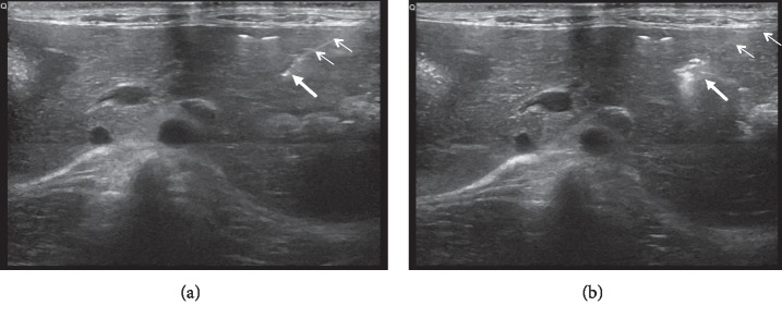
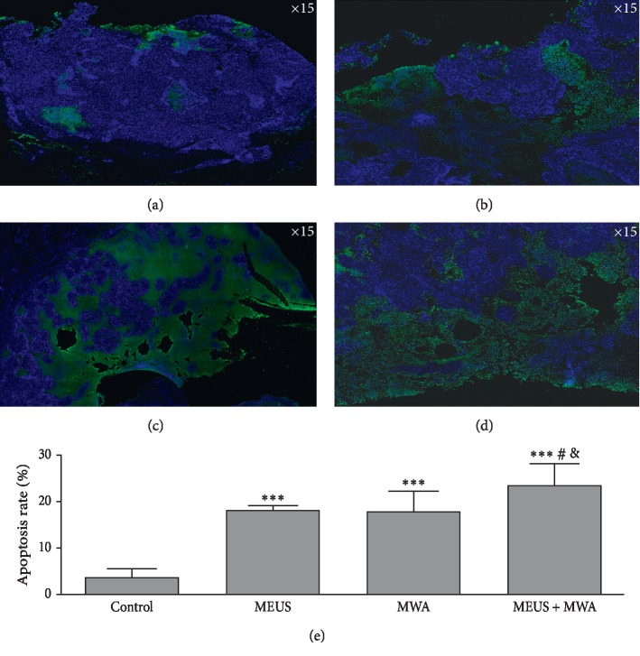
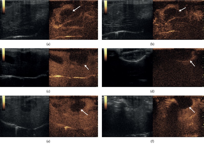
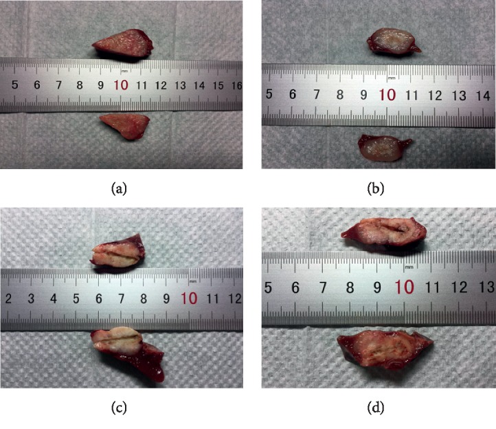
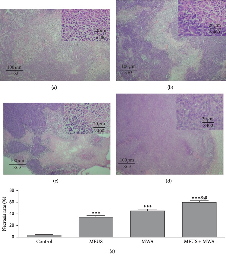
In this study, a new strategy of combining MWA with MEUS for treating HCC was proposed. Twenty-four rabbits with VX2 tumors in livers were randomly divided into MEUS + MWA, MEUS alone, MWA alone, and blank control groups, respectively ( = 6). In the MEUS group, the tumors were directly exposed to therapeutic ultrasound for 5 min with a concurrent intravenous injection of microbubbles (0.1 ml/kg diluted into 5 ml saline). In the MWA group, the tumors were treated by MWA for 1 min. In the MEUS + MWA group, tumors were ablated by MWA for 1 min after ultrasound cavitation enhanced by microbubbles as in the MEUS group. In the blank control group, the tumors received probe sham and intravenous saline. Contrast-enhanced ultrasound (CEUS) was performed before treatment and immediately after treatment to display the size, shape, and contour of the tumors. Throughout the treatment process, the local temperature of the treatment area was detected by a temperature needle punctured into the tumor. The blood samples of animals were obtained after treatment for evaluating the liver function. Tumor cell necrosis and apoptotic rates were observed after treatment by histological examination.
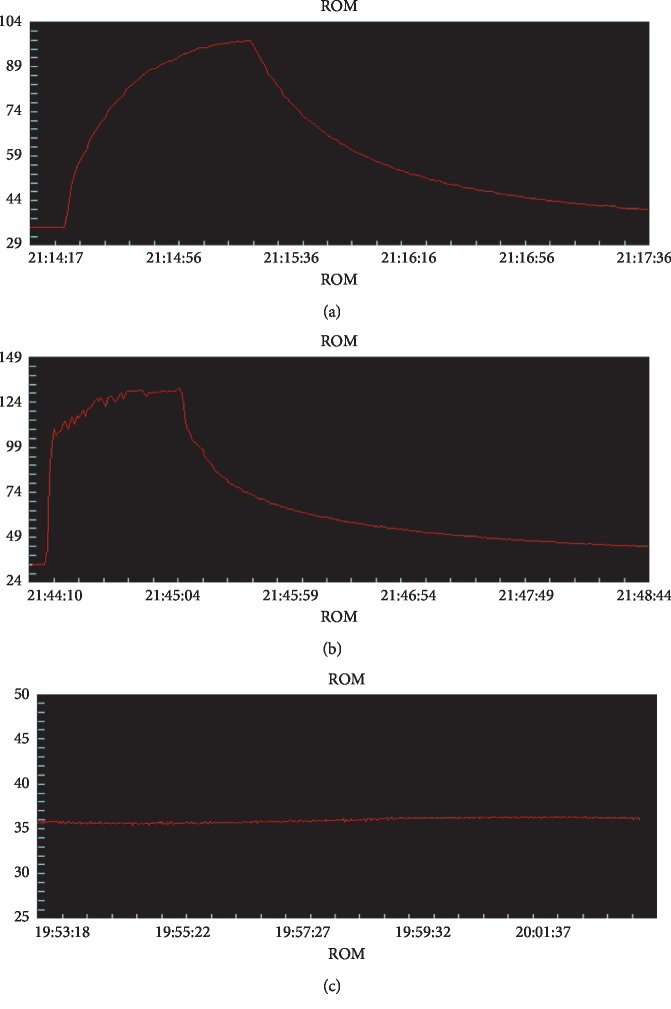
CEUS showed that although perfusion defects appeared in all the treatment groups, especially in the MEUS + MWA group, there was no significant difference between the two groups on the volumes of perfusion defects, which were 1.78 ± 0.31 (cm) in the MWA group and 1.84 ± 0.20 (cm) in the combined group ( < 0.01). The time to reach the peak temperature of the treatment area was 21.7 ± 5.0 (s) in the MWA group and 10.3 ± 5.0 (s) in the MEUS + MWA group ( < 0.01). The time to reach the peak temperature of the treatment area was 21.7 ± 5.0 (s) in the MWA group and 10.3 ± 5.0 (s) in the MEUS + MWA group ( < 0.01). The time to reach the peak temperature of the treatment area was 21.7 ± 5.0 (s) in the MWA group and 10.3 ± 5.0 (s) in the MEUS + MWA group (.
These results suggested MEUS treatment alone may significantly reduce tumor blood perfusion and led to a sharp rise in the local temperature of the treatment area to a higher PT using MEUS + MWA with higher rates of necrosis and apoptosis of cancer cells without severe liver function damage, which might be a safe strategy for treating HCC.
肝癌(HCC)等肿瘤经微波消融(MWA)治疗后复发和转移率高的原因之一是由于热沉效应,肿瘤内仍存在残留灶。微泡增强超声(MEUS)可以无创地破坏和阻断肿瘤的血液灌注,并有可能克服热沉效应,增强 MWA 的治疗效果。本研究旨在评估微泡增强超声(MEUS)在 HCC 治疗中的潜在附加益处。
本研究提出了一种将 MWA 与 MEUS 联合治疗 HCC 的新策略。24 只兔肝VX2 肿瘤随机分为 MEUS+MWA、MEUS 单独、MWA 单独和空白对照组,每组 6 只。在 MEUS 组中,直接对肿瘤进行 5 分钟的治疗超声,同时静脉注射微泡(0.1ml/kg 稀释至 5ml 生理盐水)。在 MWA 组中,肿瘤接受 MWA 治疗 1 分钟。在 MEUS+MWA 组中,在 MEUS 组超声空化增强后,用 MWA 消融 1 分钟。在空白对照组中,肿瘤接受探头假处理和静脉生理盐水。在治疗前和治疗后立即进行对比增强超声(CEUS),以显示肿瘤的大小、形状和轮廓。在整个治疗过程中,通过穿刺到肿瘤中的温度针检测治疗区域的局部温度。治疗后采集动物血液样本,评估肝功能。通过组织学检查观察治疗后肿瘤细胞坏死和凋亡率。
CEUS 显示,尽管所有治疗组均出现灌注缺损,但在 MEUS+MWA 组中更为明显,但两组灌注缺损体积无显著差异,MWA 组为 1.78±0.31(cm),联合组为 1.84±0.20(cm)(<0.01)。治疗区域达到峰值温度的时间在 MWA 组为 21.7±5.0(s),在 MEUS+MWA 组为 10.3±5.0(s)(<0.01)。治疗区域达到峰值温度的时间在 MWA 组为 21.7±5.0(s),在 MEUS+MWA 组为 10.3±5.0(s)(<0.01)。治疗区域达到峰值温度的时间在 MWA 组为 21.7±5.0(s),在 MEUS+MWA 组为 10.3±5.0(s)(<0.01)。
这些结果表明,MEUS 单独治疗可能显著降低肿瘤血液灌注,并导致 MEUS+MWA 联合治疗时治疗区域局部温度急剧升高至更高的 PT,同时伴有更高的癌细胞坏死和凋亡率,而不会造成严重的肝功能损害,这可能是治疗 HCC 的一种安全策略。