• 文献检索
  • 文档翻译
  • 深度研究
  • 学术资讯
  • Suppr Zotero 插件Zotero 插件
  • 邀请有礼
  • 套餐&价格
  • 历史记录
应用&插件
Suppr Zotero 插件Zotero 插件浏览器插件Mac 客户端Windows 客户端微信小程序
定价
高级版会员购买积分包购买API积分包
服务
文献检索文档翻译深度研究API 文档MCP 服务
关于我们
关于 Suppr公司介绍联系我们用户协议隐私条款
关注我们

Suppr 超能文献

核心技术专利:CN118964589B侵权必究
粤ICP备2023148730 号-1Suppr @ 2026

文献检索

告别复杂PubMed语法,用中文像聊天一样搜索,搜遍4000万医学文献。AI智能推荐,让科研检索更轻松。

立即免费搜索

文件翻译

保留排版,准确专业,支持PDF/Word/PPT等文件格式,支持 12+语言互译。

免费翻译文档

深度研究

AI帮你快速写综述,25分钟生成高质量综述,智能提取关键信息,辅助科研写作。

立即免费体验

计算机辅助诊断系统在超声检查甲状腺结节评估中的应用:根据放射科医生经验水平的前瞻性非劣效性研究。

Computer-Aided Diagnosis System for the Evaluation of Thyroid Nodules on Ultrasonography: Prospective Non-Inferiority Study according to the Experience Level of Radiologists.

机构信息

Department of Radiology and Research Institute of Radiology, University of Ulsan College of Medicine, Asan Medical Center, Seoul, Korea.

Department of Surgery, University of Ulsan College of Medicine, Asan Medical Center, Seoul, Korea.

出版信息

Korean J Radiol. 2020 Mar;21(3):369-376. doi: 10.3348/kjr.2019.0581.

DOI:10.3348/kjr.2019.0581
PMID:32090529
原文链接:https://pmc.ncbi.nlm.nih.gov/articles/PMC7039724/
Abstract

OBJECTIVE

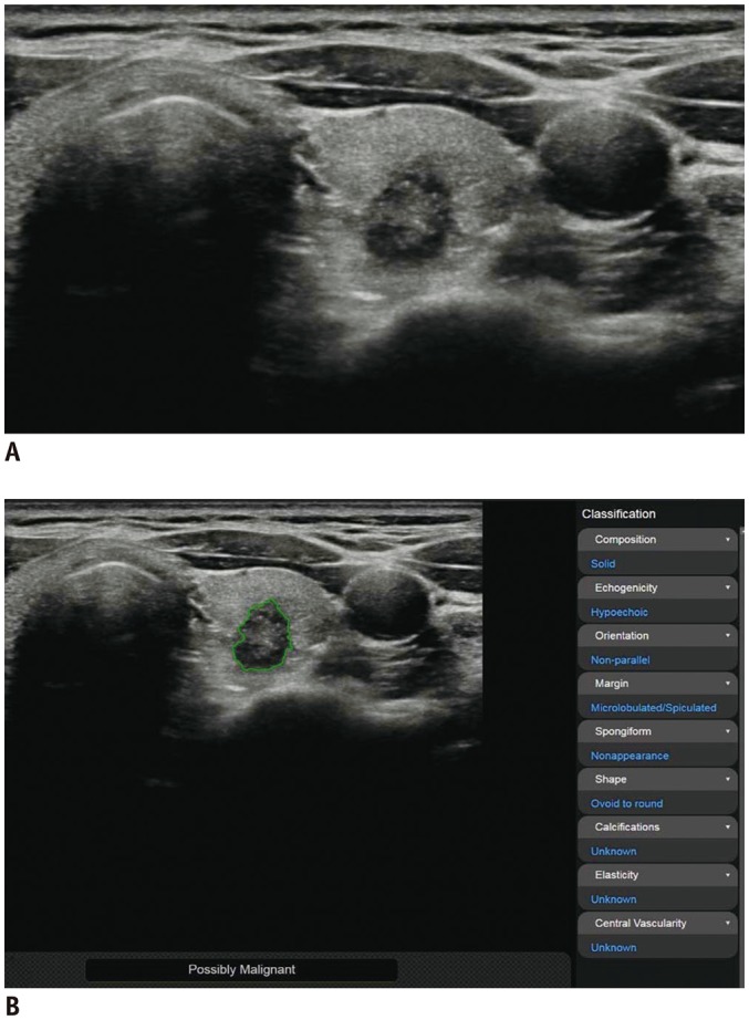
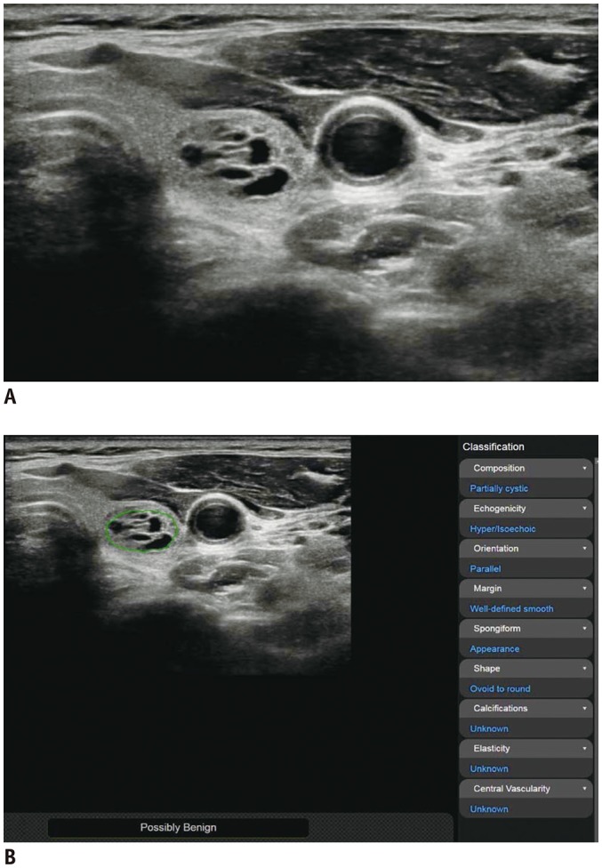
To determine whether a computer-aided diagnosis (CAD) system for the evaluation of thyroid nodules is non-inferior to radiologists with different levels of experience.

MATERIALS AND METHODS

Patients with thyroid nodules with a decisive diagnosis of benign or malignant nodule were consecutively enrolled from November 2017 to September 2018. Three radiologists with different levels of experience (1 month, 4 years, and 7 years) in thyroid ultrasound (US) reviewed the thyroid US with and without using the CAD system. Statistical analyses included non-inferiority testing of the diagnostic accuracy for malignant thyroid nodules between the CAD system and the three radiologists with a non-inferiority margin of 10%, comparison of the diagnostic performance, and the added value of the CAD system to the radiologists.

RESULTS

Altogether, 197 patients were included in the study cohort. The diagnostic accuracy of the CAD system (88.48%, 95% confidence interval [CI] = 82.65-92.53) was non-inferior to that of the radiologists with less experience (1 month and 4 year) of thyroid US (83.03%, 95% CI = 76.52-88.02; < 0.001), whereas it was inferior to that of the experienced radiologist (7 years) (95.76%, 95% CI = 91.37-97.96; = 0.138). The sensitivity and negative predictive value of the CAD system were significantly higher than those of the less-experienced radiologists were, whereas no significant difference was found with those of the experienced radiologist. A combination of US and the CAD system significantly improved sensitivity and negative predictive value, although the specificity and positive predictive value deteriorated for the less-experienced radiologists.

CONCLUSION

The CAD system may offer support for decision-making in the diagnosis of malignant thyroid nodules for operators who have less experience with thyroid US.

摘要

目的

确定计算机辅助诊断(CAD)系统在评估甲状腺结节方面是否不劣于不同经验水平的放射科医生。

材料与方法

从 2017 年 11 月至 2018 年 9 月连续纳入具有明确良性或恶性结节诊断的甲状腺结节患者。3 名具有不同甲状腺超声(US)经验水平(1 个月、4 年和 7 年)的放射科医生在使用和不使用 CAD 系统的情况下对甲状腺 US 进行了回顾。统计分析包括 CAD 系统与经验较少的 3 名放射科医生(非劣效性边界为 10%)对恶性甲状腺结节的诊断准确性的非劣效性检验、诊断性能比较以及 CAD 系统对放射科医生的增值。

结果

共有 197 名患者纳入研究队列。CAD 系统的诊断准确性(88.48%,95%置信区间[CI] = 82.65-92.53)不劣于经验较少的甲状腺 US 放射科医生(1 个月和 4 年)(83.03%,95% CI = 76.52-88.02;<0.001),但劣于经验丰富的放射科医生(7 年)(95.76%,95% CI = 91.37-97.96;=0.138)。CAD 系统的敏感性和阴性预测值显著高于经验较少的放射科医生,而与经验丰富的放射科医生无显著差异。US 与 CAD 系统的联合应用显著提高了敏感性和阴性预测值,尽管经验较少的放射科医生的特异性和阳性预测值有所恶化。

结论

对于甲状腺 US 经验较少的操作人员,CAD 系统可为恶性甲状腺结节的诊断决策提供支持。

https://cdn.ncbi.nlm.nih.gov/pmc/blobs/9626/7039724/efde54cbdd51/kjr-21-369-g002.jpg
https://cdn.ncbi.nlm.nih.gov/pmc/blobs/9626/7039724/2e37a05fb583/kjr-21-369-g001.jpg
https://cdn.ncbi.nlm.nih.gov/pmc/blobs/9626/7039724/efde54cbdd51/kjr-21-369-g002.jpg
https://cdn.ncbi.nlm.nih.gov/pmc/blobs/9626/7039724/2e37a05fb583/kjr-21-369-g001.jpg
https://cdn.ncbi.nlm.nih.gov/pmc/blobs/9626/7039724/efde54cbdd51/kjr-21-369-g002.jpg

相似文献

1
Computer-Aided Diagnosis System for the Evaluation of Thyroid Nodules on Ultrasonography: Prospective Non-Inferiority Study according to the Experience Level of Radiologists.计算机辅助诊断系统在超声检查甲状腺结节评估中的应用:根据放射科医生经验水平的前瞻性非劣效性研究。
Korean J Radiol. 2020 Mar;21(3):369-376. doi: 10.3348/kjr.2019.0581.
2
Computer-aided diagnosis system for thyroid nodules on ultrasonography: diagnostic performance and reproducibility based on the experience level of operators.基于操作人员经验水平的超声甲状腺结节计算机辅助诊断系统:诊断性能和可重复性。
Eur Radiol. 2019 Apr;29(4):1978-1985. doi: 10.1007/s00330-018-5772-9. Epub 2018 Oct 22.
3
A Computer-Aided Diagnosis System Using Artificial Intelligence for the Diagnosis and Characterization of Thyroid Nodules on Ultrasound: Initial Clinical Assessment.一种使用人工智能的计算机辅助诊断系统,用于超声检查中甲状腺结节的诊断与特征描述:初步临床评估
Thyroid. 2017 Apr;27(4):546-552. doi: 10.1089/thy.2016.0372. Epub 2017 Feb 28.
4
Ultrasound Computer-Aided Diagnosis (CAD) Based on the Thyroid Imaging Reporting and Data System (TI-RADS) to Distinguish Benign from Malignant Thyroid Nodules and the Diagnostic Performance of Radiologists with Different Diagnostic Experience.基于甲状腺影像报告和数据系统 (TI-RADS) 的超声计算机辅助诊断 (CAD) 对甲状腺良恶性结节的鉴别诊断及不同诊断经验的放射科医生的诊断效能
Med Sci Monit. 2020 Jan 2;26:e918452. doi: 10.12659/MSM.918452.
5
Effectiveness evaluation of computer-aided diagnosis system for the diagnosis of thyroid nodules on ultrasound: A systematic review and meta-analysis.超声甲状腺结节诊断中计算机辅助诊断系统的有效性评估:一项系统评价和荟萃分析
Medicine (Baltimore). 2019 Aug;98(32):e16379. doi: 10.1097/MD.0000000000016379.
6
Computer-aided diagnosis system of thyroid nodules ultrasonography: Diagnostic performance difference between computer-aided diagnosis and 111 radiologists.甲状腺结节超声检查的计算机辅助诊断系统:计算机辅助诊断与111名放射科医生之间的诊断性能差异
Medicine (Baltimore). 2020 Jun 5;99(23):e20634. doi: 10.1097/MD.0000000000020634.
7
Computer-Aided Diagnosis of Thyroid Nodules via Ultrasonography: Initial Clinical Experience.计算机辅助超声诊断甲状腺结节:初步临床经验。
Korean J Radiol. 2018 Jul-Aug;19(4):665-672. doi: 10.3348/kjr.2018.19.4.665. Epub 2018 Jun 14.
8
Computer-aided diagnosis for classifying benign versus malignant thyroid nodules based on ultrasound images: A comparison with radiologist-based assessments.基于超声图像的甲状腺良恶性结节分类的计算机辅助诊断:与放射科医生评估的比较。
Med Phys. 2016 Jan;43(1):554. doi: 10.1118/1.4939060.
9
A computer-aided diagnosing system in the evaluation of thyroid nodules-experience in a specialized thyroid center.计算机辅助诊断系统在甲状腺结节评估中的应用——在一个甲状腺专科中心的经验。
World J Surg Oncol. 2019 Dec 6;17(1):210. doi: 10.1186/s12957-019-1752-z.
10
A computer-aided diagnosis system for the assessment and characterization of low-to-high suspicion thyroid nodules on ultrasound.用于超声评估和诊断低-高度疑似甲状腺结节的计算机辅助诊断系统。
Radiol Med. 2019 Feb;124(2):118-125. doi: 10.1007/s11547-018-0942-z. Epub 2018 Sep 22.

引用本文的文献

1
Enhancing diagnostic precision for thyroid C-TIRADS category 4 nodules: a hybrid deep learning and machine learning model integrating grayscale and elastographic ultrasound features.提高甲状腺C-TIRADS 4类结节的诊断准确性:一种整合灰阶和弹性成像超声特征的深度学习与机器学习混合模型
Quant Imaging Med Surg. 2025 Sep 1;15(9):7951-7963. doi: 10.21037/qims-2025-594. Epub 2025 Aug 11.
2
Multiparametric ultrasound techniques are superior to AI-assisted ultrasound for assessment of solid thyroid nodules: a prospective study.多参数超声技术在评估实性甲状腺结节方面优于人工智能辅助超声:一项前瞻性研究。
Endocrine. 2025 Jul 10. doi: 10.1007/s12020-025-04306-8.
3

本文引用的文献

1
Effectiveness evaluation of computer-aided diagnosis system for the diagnosis of thyroid nodules on ultrasound: A systematic review and meta-analysis.超声甲状腺结节诊断中计算机辅助诊断系统的有效性评估:一项系统评价和荟萃分析
Medicine (Baltimore). 2019 Aug;98(32):e16379. doi: 10.1097/MD.0000000000016379.
2
Real-World Performance of Computer-Aided Diagnosis System for Thyroid Nodules Using Ultrasonography.基于超声的计算机辅助诊断系统在甲状腺结节中的真实世界性能。
Ultrasound Med Biol. 2019 Oct;45(10):2672-2678. doi: 10.1016/j.ultrasmedbio.2019.05.032. Epub 2019 Jun 29.
3
Computer-aided diagnosis system for thyroid nodules on ultrasonography: diagnostic performance and reproducibility based on the experience level of operators.
Ultrasonic radiomics in predicting pathologic type for thyroid cancer: a preliminary study using radiomics features for predicting medullary thyroid carcinoma.
超声影像组学在预测甲状腺癌病理类型中的应用:一项利用影像组学特征预测甲状腺髓样癌的初步研究
Front Endocrinol (Lausanne). 2025 Feb 19;16:1428888. doi: 10.3389/fendo.2025.1428888. eCollection 2025.
4
Human-AI collaboration for ultrasound diagnosis of thyroid nodules: a clinical trial.人机协作用于甲状腺结节的超声诊断:一项临床试验
Eur Arch Otorhinolaryngol. 2025 Feb 8. doi: 10.1007/s00405-025-09236-9.
5
Improving the diagnostic performance of inexperienced readers for thyroid nodules through digital self-learning and artificial intelligence assistance.通过数字自学和人工智能辅助提高经验不足的读者对甲状腺结节的诊断能力。
Front Endocrinol (Lausanne). 2024 Jul 2;15:1372397. doi: 10.3389/fendo.2024.1372397. eCollection 2024.
6
Artificial intelligence in thyroid ultrasound.甲状腺超声中的人工智能
Front Oncol. 2023 May 12;13:1060702. doi: 10.3389/fonc.2023.1060702. eCollection 2023.
7
Comparison of S-Detect and thyroid imaging reporting and data system classifications in the diagnosis of cytologically indeterminate thyroid nodules.S-Detect 与甲状腺影像报告和数据系统分类在诊断细胞学不确定的甲状腺结节中的比较。
Front Endocrinol (Lausanne). 2023 Jan 24;14:1098031. doi: 10.3389/fendo.2023.1098031. eCollection 2023.
8
Investigating the diagnostic efficiency of a computer-aided diagnosis system for thyroid nodules in the context of Hashimoto's thyroiditis.在桥本甲状腺炎背景下研究计算机辅助诊断系统对甲状腺结节的诊断效率。
Front Oncol. 2023 Jan 5;12:941673. doi: 10.3389/fonc.2022.941673. eCollection 2022.
9
Clinical value of artificial intelligence in thyroid ultrasound: a prospective study from the real world.人工智能在甲状腺超声中的临床价值:来自真实世界的前瞻性研究。
Eur Radiol. 2023 Jul;33(7):4513-4523. doi: 10.1007/s00330-022-09378-y. Epub 2023 Jan 9.
10
Artificial Intelligence for Pre-operative Diagnosis of Malignant Thyroid Nodules Based on Sonographic Features and Cytology Category.基于超声特征和细胞学分类的甲状腺恶性结节术前人工智能诊断。
World J Surg. 2023 Feb;47(2):330-339. doi: 10.1007/s00268-022-06798-1. Epub 2022 Nov 6.
基于操作人员经验水平的超声甲状腺结节计算机辅助诊断系统:诊断性能和可重复性。
Eur Radiol. 2019 Apr;29(4):1978-1985. doi: 10.1007/s00330-018-5772-9. Epub 2018 Oct 22.
4
A computer-aided diagnosis system for the assessment and characterization of low-to-high suspicion thyroid nodules on ultrasound.用于超声评估和诊断低-高度疑似甲状腺结节的计算机辅助诊断系统。
Radiol Med. 2019 Feb;124(2):118-125. doi: 10.1007/s11547-018-0942-z. Epub 2018 Sep 22.
5
Computer-Aided Diagnosis of Thyroid Nodules via Ultrasonography: Initial Clinical Experience.计算机辅助超声诊断甲状腺结节:初步临床经验。
Korean J Radiol. 2018 Jul-Aug;19(4):665-672. doi: 10.3348/kjr.2018.19.4.665. Epub 2018 Jun 14.
6
Computer-aided system for diagnosing thyroid nodules on ultrasound: A comparison with radiologist-based clinical assessments.用于超声诊断甲状腺结节的计算机辅助系统:与基于放射科医生的临床评估的比较。
Head Neck. 2018 Apr;40(4):778-783. doi: 10.1002/hed.25049. Epub 2017 Dec 29.
7
Computer-aided diagnosis of malignant or benign thyroid nodes based on ultrasound images.基于超声图像的甲状腺良恶性结节的计算机辅助诊断
Eur Arch Otorhinolaryngol. 2017 Jul;274(7):2891-2897. doi: 10.1007/s00405-017-4562-3. Epub 2017 Apr 7.
8
A Computer-Aided Diagnosis System Using Artificial Intelligence for the Diagnosis and Characterization of Thyroid Nodules on Ultrasound: Initial Clinical Assessment.一种使用人工智能的计算机辅助诊断系统,用于超声检查中甲状腺结节的诊断与特征描述:初步临床评估
Thyroid. 2017 Apr;27(4):546-552. doi: 10.1089/thy.2016.0372. Epub 2017 Feb 28.
9
Computer-aided diagnosis for classifying benign versus malignant thyroid nodules based on ultrasound images: A comparison with radiologist-based assessments.基于超声图像的甲状腺良恶性结节分类的计算机辅助诊断:与放射科医生评估的比较。
Med Phys. 2016 Jan;43(1):554. doi: 10.1118/1.4939060.
10
2015 American Thyroid Association Management Guidelines for Adult Patients with Thyroid Nodules and Differentiated Thyroid Cancer: The American Thyroid Association Guidelines Task Force on Thyroid Nodules and Differentiated Thyroid Cancer.2015年美国甲状腺协会成人甲状腺结节和分化型甲状腺癌管理指南:美国甲状腺协会甲状腺结节和分化型甲状腺癌指南工作组
Thyroid. 2016 Jan;26(1):1-133. doi: 10.1089/thy.2015.0020.