Jamet Bastien, Bailly Clément, Carlier Thomas, Touzeau Cyrille, Michaud Anne-Victoire, Bourgeois Mickael, Moreau Philippe, Bodet-Milin Caroline, Kraeber-Bodere Françoise
Nuclear Medicine Unit, University Hospital, 44093 Nantes, France.
CRCINA, INSERM, CNRS, Angers University, Nantes University, 44093 Nantes, France.
Cancers (Basel). 2020 Feb 19;12(2):486. doi: 10.3390/cancers12020486.
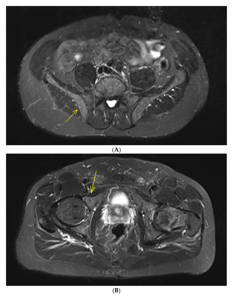
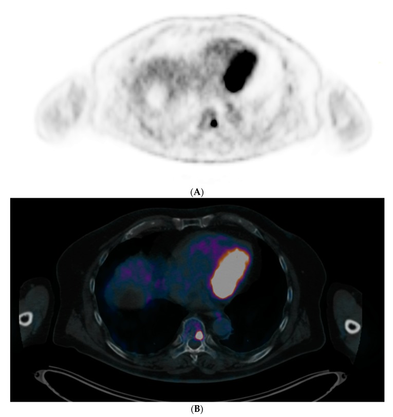
Multiple myeloma (MM) is always preceded by an initial monoclonal gammopathy of undetermined significance (MGUS) that then develops into asymptomatic or smoldering multiple myeloma (SMM), which constitutes an intermediate clinical stage between MGUS and MM. According to a recent study, risk factors for faster MGUS to MM progression include an M protein of 1.5 g/dL or more and an abnormal free light chain ratio in patients with non-IgM MGUS. Therefore, the International Myeloma Working Group (IMWG) decided to recommend whole-body computed tomography (WBCT) for patients with high-risk MGUS in order to exclude early bone destruction. Studies evaluating magnetic resonance imaging (MRI) in SMM found an optimal cutoff of two or more focal lesions to be of prognostic significance for fast progression into symptomatic disease and considered this biomarker as a myeloma-defining event (MDE) needing to start therapy with the aim to avoid progression to harmful bone lesions. Moreover, studies assessing positron emission tomography (PET) with computed tomography (CT) using 18F-deoxyglucose (FDG) (FDG-PET/CT) in SMM showed that presence of focal bone lesion without underlying osteolysis is associated with a rapid progression to symptomatic MM. Latest IMWG guidelines recommended to perform WBCT (either CT alone or as part of an FDG-PET/CT protocol) as the first imaging technique at suspected SMM and, if these images are negative or inconclusive, to perform whole-body MRI. The goal of this paper is to clarify the role of different imaging modalities in MGUS and SMM workups.
多发性骨髓瘤(MM)之前通常会有意义未明的单克隆丙种球蛋白病(MGUS),随后发展为无症状或冒烟型多发性骨髓瘤(SMM),SMM构成了MGUS和MM之间的一个中间临床阶段。根据最近的一项研究,MGUS向MM快速进展的危险因素包括非IgM型MGUS患者的M蛋白≥1.5 g/dL以及游离轻链比例异常。因此,国际骨髓瘤工作组(IMWG)决定建议对高危MGUS患者进行全身计算机断层扫描(WBCT),以排除早期骨质破坏。评估SMM中磁共振成像(MRI)的研究发现,两个或更多局灶性病变的最佳临界值对快速进展为有症状疾病具有预后意义,并将该生物标志物视为需要开始治疗以避免进展为有害骨病变的骨髓瘤定义事件(MDE)。此外,评估SMM中使用18F-脱氧葡萄糖(FDG)的正电子发射断层扫描(PET)与计算机断层扫描(CT)(FDG-PET/CT)的研究表明,无潜在骨质溶解的局灶性骨病变与快速进展为有症状的MM相关。IMWG最新指南建议,在疑似SMM时,将WBCT(单独的CT或作为FDG-PET/CT方案的一部分)作为首选成像技术,如果这些图像为阴性或不确定,则进行全身MRI。本文的目的是阐明不同成像方式在MGUS和SMM检查中的作用。