Hellinen Laura, Hongisto Heidi, Ramsay Eva, Kaarniranta Kai, Vellonen Kati-Sisko, Skottman Heli, Ruponen Marika
School of Pharmacy, Faculty of Health Sciences, University of Eastern Finland, 70210 Kuopio, Finland.
Department of Ophthalmology, Institute of Clinical Medicine, University of Eastern Finland, 70210 Kuopio, Finland.
Pharmaceutics. 2020 Feb 19;12(2):176. doi: 10.3390/pharmaceutics12020176.
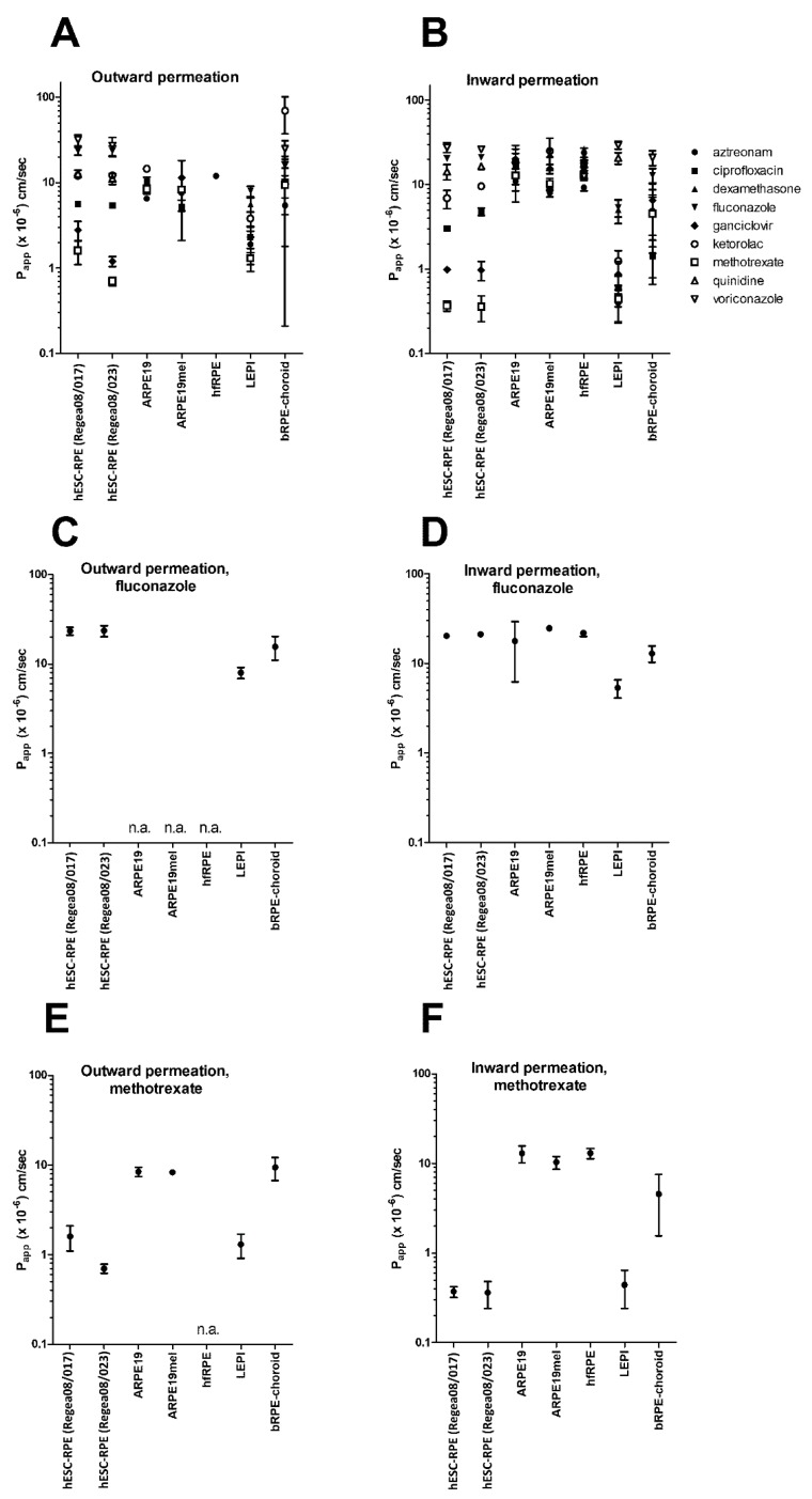
The retinal pigment epithelial (RPE) cell monolayer forms the outer blood-retinal barrier and has a crucial role in ocular pharmacokinetics. Although several RPE cell models are available, there have been no systematic comparisons of their barrier properties with respect to drug permeability. We compared the barrier properties of several RPE secondary cell lines (ARPE19, ARPE19mel, and LEPI) and both primary (hfRPE) and stem-cell derived RPE (hESC-RPE) cells by investigating the permeability of nine drugs (aztreonam, ciprofloxacin, dexamethasone, fluconazole, ganciclovir, ketorolac, methotrexate, voriconazole, and quinidine) across cell monolayers. ARPE19, ARPE19mel, and hfRPE cells displayed a narrow P value range, with relatively high permeation rates (5.2-26 × 10 cm/s. In contrast, hESC-RPE and LEPI cells efficiently restricted the drug flux, and displayed even lower P values than those reported for bovine RPE-choroid, with the range of 0.4-32 cm/s (hESC-RPE cells) and 0.4-29 × 10 cm/s, (LEPI cells). Therefore, ARPE19, ARPE19mel, and hfRPE cells failed to form a tight barrier, whereas hESC-RPE and LEPI cells restricted the drug flux to a similar extent as bovine RPE-choroid. Therefore, LEPI and hESC-RPE cells are valuable tools in ocular drug discovery.
视网膜色素上皮(RPE)细胞单层构成了外血视网膜屏障,在眼部药代动力学中起着关键作用。尽管有几种RPE细胞模型可用,但尚未对它们在药物通透性方面的屏障特性进行系统比较。我们通过研究九种药物(氨曲南、环丙沙星、地塞米松、氟康唑、更昔洛韦、酮咯酸、甲氨蝶呤、伏立康唑和奎尼丁)跨细胞单层的通透性,比较了几种RPE传代细胞系(ARPE19、ARPE19mel和LEPI)以及原代(hfRPE)和干细胞衍生的RPE(hESC-RPE)细胞的屏障特性。ARPE19、ARPE19mel和hfRPE细胞显示出较窄的P值范围,具有相对较高的渗透速率(5.2 - 26×10 cm/s)。相比之下,hESC-RPE和LEPI细胞有效地限制了药物通量,并且显示出比报道的牛RPE - 脉络膜更低的P值,范围为0.4 - 32 cm/s(hESC-RPE细胞)和0.4 - 29×10 cm/s(LEPI细胞)。因此,ARPE19、ARPE19mel和hfRPE细胞未能形成紧密屏障,而hESC-RPE和LEPI细胞限制药物通量的程度与牛RPE - 脉络膜相似。因此,LEPI和hESC-RPE细胞是眼部药物研发中有价值的工具。