Cavalheiro Cristina Schmitt, Arcuri Marcel Henrique, Guil Victor Reis, Gali Julio Cesar
Pontifícia Universidade Católica de São Paulo, School of Medical Sciences and Health, Graduate Training Program in Orthopedics and Traumatology, Sorocaba, SP, Brazil.
Pontifícia Universidade Católica de São Paulo, School of Medical Sciences and Health, Department of Surgery, Sorocaba, SP, Brazil.
Acta Ortop Bras. 2020 Jan-Feb;28(1):12-15. doi: 10.1590/1413-785220202801226897.
To describe the anatomical and pathological osteoarticular, muscular and tendinous variations in feet of cadavers with hallux valgus and to correlate them with the degree of radiographic deformity.
Dissections and radiographs were conducted in the feet of 22 cadavers with halux valgus, aged between 20 and 70 years. The feet affected were compared with 5 normal feet in order to document the anatomical and pathological, myotendinous and articular variations found.
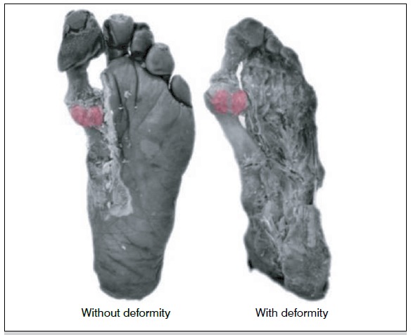
The extensor hallucis longus and brevis tendons were arched in all degrees of deformity, causing a lateral deviation that forms the arc chord of the metatarsophalangeal angle of the hallux. We also observed a deviation to the plantar face of the abductor muscle tendon and lateral deviation of the flexor hallucis muscle tendon. In the moderate deformities, the medial deviation of the first metatarsal head was observed, sliding out of the sesamoid apparatus, pronation of this head, and formation of medial exostoses. In severe deformities, in addition to all other deformities, we found the extensor hallucis longus tendon with two distal insertions, rather than just one.
The anatomical alterations found in the hallux valgus may be related to the degree of radiographic deformity
描述患有拇外翻的尸体足部的解剖学和病理学上的骨关节、肌肉和肌腱变异,并将它们与影像学畸形程度相关联。
对22具年龄在20至70岁之间患有拇外翻的尸体足部进行解剖和X光摄影。将患足与5只正常足部进行比较,以记录所发现的解剖学和病理学、肌腱和关节变异。
在所有畸形程度中,拇长伸肌腱和拇短伸肌腱呈弓形,导致外侧偏移,形成拇趾跖趾角的弧形弦。我们还观察到拇展肌肌腱向足底面偏移以及拇长屈肌肌腱外侧偏移。在中度畸形中,观察到第一跖骨头向内侧偏移,滑出籽骨装置,该跖骨头旋前,并形成内侧骨赘。在重度畸形中,除了所有其他畸形外,我们发现拇长伸肌腱有两个远端附着点,而不是只有一个。
拇外翻中发现的解剖学改变可能与影像学畸形程度有关。