,.
Invest Ophthalmol Vis Sci. 2020 Feb 7;61(2):40. doi: 10.1167/iovs.61.2.40.
To determine the relationship between funduscopic findings in myopic eyes and the prevalence and structure of the conus in the optical coherence tomographic (OCT) images.
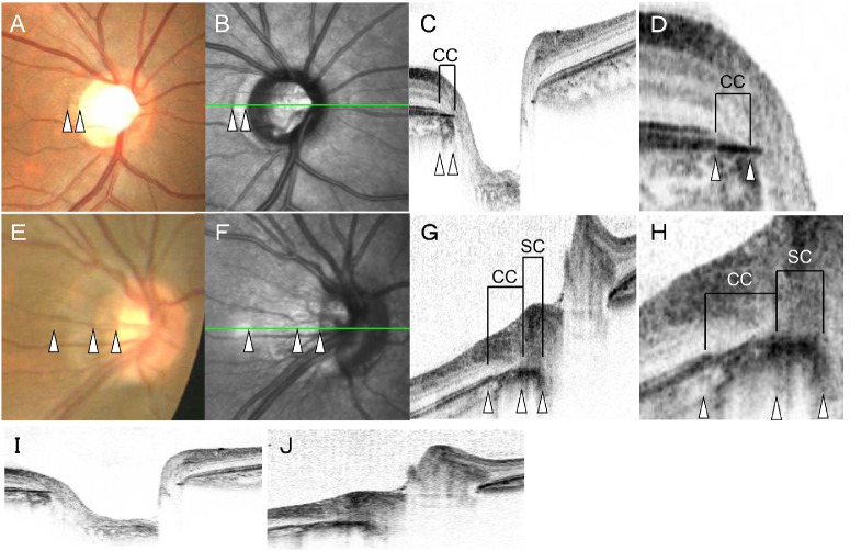
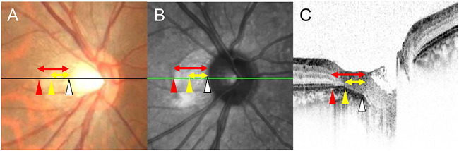
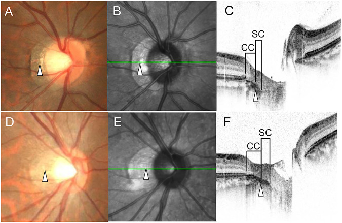
A prospective observational cross-sectional study of 121 right eyes of 121 young healthy volunteers. All participants underwent color fundus photography (CFP), scanning laser ophthalmoscopy, and OCT. Based on the OCT analyses, the area between the edge of the ellipsoid zone (EZ) and that of choroid was defined as the "choroidal conus (CC)", and the area between the edge of the choroid and the scleral edge as the "scleral conus (SC)". The eyes were classified into three groups such as the non-conus (NC) group, CC group, and SC group. The differences in the axial length, optic disc tilt, ovality ratio, papillomacular position angle, and peripapillary nerve fiber elevation (pNFE) between the three groups were determined.
CFPs detected a conus in 79 eyes (65.3 %). The outer border of the conus in CFPs corresponded with the edge of the EZ in the OCT in all subjects. Thirty-seven eyes had CC alone (CC group) and 42 eyes had both CC and SC (SC group). The CC and SC groups had longer axial lengths and more frequent pNFEs than the NC group. There was a significant difference in the optic disc tilt and ovality ratio between the CC and SC groups.
The eyes with SC tend to have larger optic disc tilt and smaller ovality ratio than the eyes with CC only.
确定近视眼中眼底表现与光学相干断层扫描(OCT)图像中圆锥体的患病率和结构之间的关系。
对 121 名年轻健康志愿者的 121 只右眼进行前瞻性观察性横断面研究。所有参与者均接受了眼底彩色照相(CFP)、扫描激光检眼镜和 OCT 检查。根据 OCT 分析,将椭圆体带(EZ)边缘与脉络膜边缘之间的区域定义为“脉络膜圆锥(CC)”,脉络膜边缘与巩膜边缘之间的区域定义为“巩膜圆锥(SC)”。将眼睛分为非圆锥体(NC)组、CC 组和 SC 组。比较三组间眼轴长度、视盘倾斜度、椭圆度比、乳头黄斑位置角和神经纤维层旁隆起(pNFE)的差异。
CFP 检测到 79 只眼(65.3%)有圆锥体。所有受试者的圆锥体 CFPs 外边界与 OCT 中的 EZ 边缘相对应。37 只眼仅有 CC(CC 组),42 只眼同时有 CC 和 SC(SC 组)。CC 组和 SC 组的眼轴长度较长,pNFE 较常见。CC 组和 SC 组之间的视盘倾斜度和椭圆度比存在显著差异。
与仅存在 CC 的眼相比,存在 SC 的眼的视盘倾斜度更大,椭圆度比更小。