The Affiliated Eye Hospital of Wenzhou Medical University, Hangzhou, Zhejiang, China.
Department of Ophthalmology, Zhejiang Provincial People's Hospital, Hanghzou, Zhejiang, China.
Invest Ophthalmol Vis Sci. 2022 Jun 1;63(6):20. doi: 10.1167/iovs.63.6.20.
To investigate the characteristics of the optic nerve head (ONH) in myopia using swept-source optical coherence tomography (SS-OCT).
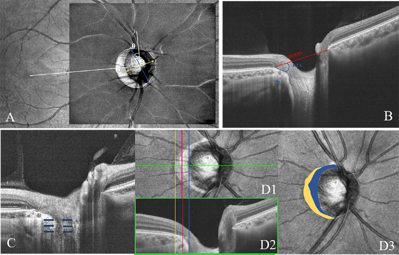
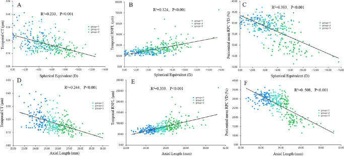
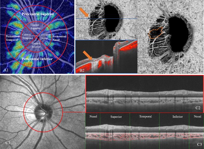
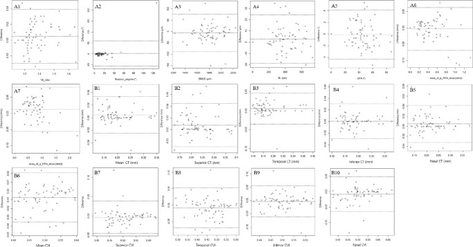
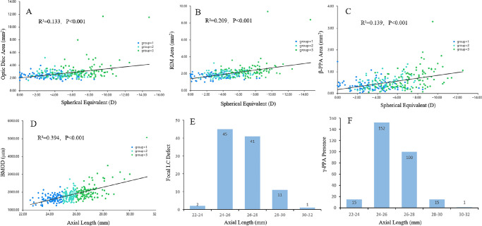
Participants were divided into three groups according to the axial length (AL). The optic disc morphology, retinal nerve fiber layer (RNFL) thickness, and radial peripapillary capillary (RPC) vessel density (VD), optic disc tilt, rotation, Bruch's membrane opening distance (BMOD), border length (BL), border tissue angle, focal lamina cribrosa (LC) defects, β- and γ-zone peripapillary atrophy (PPA), microvasculature dropout (MvD), choroidal thickness (CT), and the choroidal vascularity index (CVI) were compared. Linear regression analysis evaluated relationships between spherical equivalent, AL, and ONH parameters.
One hundred five, 98, and 118 eyes were included in groups 1, 2, and 3, respectively. With AL increasing, the mean, superior and temporal CT, central mean and temporal, pericentral mean, inferior and nasal RPC VD, and temporal CVI decreased, whereas the mean and temporal RNFL thickness, optic disc, RIM and β-PPA area, presence and area of γ-PPA, BMOD and BL increased. Compared to other groups, group 3 depicted a larger cup area, more focal LC defect and total and juxtapapillary MvD; a lower central superior, inferior and nasal, pericentral superior, and temporal RPC VD. Group 1 demonstrated more tilted disc, larger inferior and nasal CT, mean, superior, inferior, and nasal CVI.
Myopia eyes have larger ONH changes, PPAs, regional RNFL, and MvD, but smaller regional CTs, RPC VD, and CVIs. SS-OCT may be useful in detecting ONH variations during myopia.
利用扫频源光学相干断层扫描(SS-OCT)研究近视患者视神经头(ONH)的特征。
根据眼轴(AL)将参与者分为三组。比较视盘形态、视网膜神经纤维层(RNFL)厚度、放射状神经纤维层(RPC)血管密度(VD)、视盘倾斜、旋转、Bruch 膜开口距离(BMOD)、边界长度(BL)、边界组织角度、焦点层状筛板(LC)缺损、β-和γ-区视盘周围萎缩(PPA)、微血管缺失(MvD)、脉络膜厚度(CT)和脉络膜血管指数(CVI)。线性回归分析评估了球镜等效、AL 和 ONH 参数之间的关系。
分别有 105、98 和 118 只眼纳入第 1、2 和 3 组。随着 AL 的增加,平均、上和颞侧 CT、中央平均和颞侧、中心旁平均、下和鼻侧 RPC VD、以及颞侧 CVI 降低,而平均和颞侧 RNFL 厚度、视盘、RIM 和β-PPA 面积、γ-PPA 的存在和面积、BMOD 和 BL 增加。与其他组相比,第 3 组杯面积更大,焦点 LC 缺损和总、近旁 MvD 更多;中央上、下和鼻侧、中心旁上、颞侧 RPC VD 更低。第 1 组视盘倾斜度更大,下和鼻侧 CT、平均、上、下和鼻侧 CVI 更大。
近视眼中 ONH 变化、PPA、区域性 RNFL 和 MvD 更大,但区域性 CT、RPC VD 和 CVI 更小。SS-OCT 可能有助于检测近视中 ONH 的变化。