Department of Radiology and Nuclear Medicine, Amsterdam University Medical Center, Location VUMC, Amsterdam, The Netherlands.
Department of Biomedical Engineering, Institute Hall, Rochester Institute of Technology (RIT), Rochester, New York, NY, United States of America.
PLoS One. 2020 Feb 26;15(2):e0229444. doi: 10.1371/journal.pone.0229444. eCollection 2020.
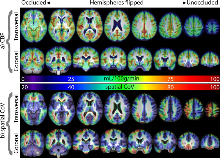
Clinical interpretation of arterial spin labeling (ASL) perfusion MRI in cerebrovascular disease remains challenging mainly because of the method's sensitivity to concomitant contributions from both intravascular and tissue compartments. While acquisition of multi-delay images can differentiate between the two contributions, the prolonged acquisition is prone to artifacts and not practical for clinical applications. Here, the utility of the spatial coefficient of variation (sCoV) of a single-delay ASL image as a marker of the intravascular contribution was evaluated by testing the hypothesis that sCoV can detect the effects of differences in label arrival times between ipsi- and contra-lateral hemispheres even in the absence of a hemispheric difference in CBF. Hemispheric lateralization values for sCoV and CBF were computed from ASL images acquired on 28 patients (age 73.9 ± 10.2 years, 8 women) with asymptomatic unilateral carotid occlusion. The results showed that sCoV lateralization predicted the occluded side with 96.4% sensitivity, missing only 1 patient. In contrast, the sensitivity of the CBF lateralization was 71.4%, with 8 patients showing no difference in CBF between hemispheres. The findings demonstrate the potential clinical utility of sCoV as a cerebrovascular correlate of large vessel disease. Using sCoV in tandem with CBF, vascular information can be obtained in image processing without the need for additional scan-time.
临床解读脑血管疾病的动脉自旋标记(ASL)灌注 MRI 仍然具有挑战性,主要是因为该方法对血管内和组织容积两者的贡献均很敏感。尽管采集多延迟图像可以区分这两种贡献,但延长采集时间容易产生伪影,不适合临床应用。在这里,通过检验以下假设评估了单延迟 ASL 图像空间变异系数(sCoV)作为血管内贡献标记的效用:sCoV 可以检测到即使在 CBF 没有半球差异的情况下,来自对侧和同侧半球的标记到达时间的差异的影响。使用从 28 例无症状单侧颈动脉闭塞患者(年龄 73.9 ± 10.2 岁,8 名女性)获得的 ASL 图像计算了 sCoV 和 CBF 的半球侧化值。结果表明,sCoV 侧化以 96.4%的敏感性预测了闭塞侧,仅漏诊 1 例患者。相比之下,CBF 侧化的敏感性为 71.4%,8 例患者的半球间 CBF 无差异。这些发现表明 sCoV 作为大血管疾病的脑血管相关物具有潜在的临床应用价值。在图像处理中,sCoV 与 CBF 联合使用,可以在无需额外扫描时间的情况下获得血管信息。