Tyrtyshnaia Anna, Manzhulo Igor
A.V. Zhirmunsky National Scientific Center of Marine Biology, Far Eastern Branch, Russian Academy of Sciences, Vladivostok, Russia.
J Pain Res. 2020 Feb 11;13:345-354. doi: 10.2147/JPR.S238458. eCollection 2020.
Neuropathic pain manifests in a diverse combination of sensory symptoms and disorders of higher nervous activity, such as memory deficiency, anxiety, depression, anhedonia, etc. This suggests the participation of brain structures, including the hippocampus, in the pathogenesis of neuropathic pain. The elucidation of central sensitization mechanisms underlying neuropathic pain cognitive and affective symptoms may be useful in the development of new and effective treatments for these common disorders. The study aims to elucidate the effect of chronic neuropathic pain on cognitive function and underlying neuronal plasticity in the hippocampus.
Chronic constriction injury of mouse right hind limb sciatic nerve was used as a model of neuropathic pain. The presence of neuropathic pain was confirmed by the thermal and mechanical allodynia. The morphology of the CA1 pyramidal neurons and the dentate gyrus (DG) granule neurons were studied using Golgi-Cox staining. The hippocampal proteins concentration was determined by immunohistochemistry and ELISA.
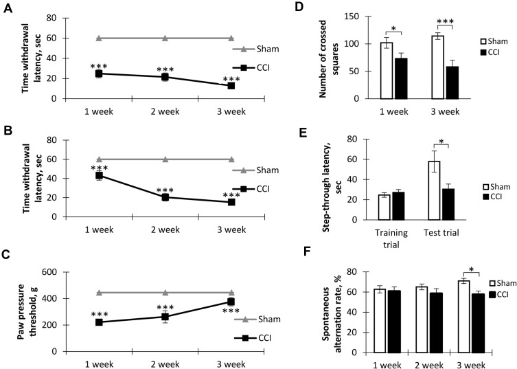
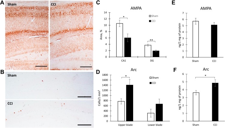
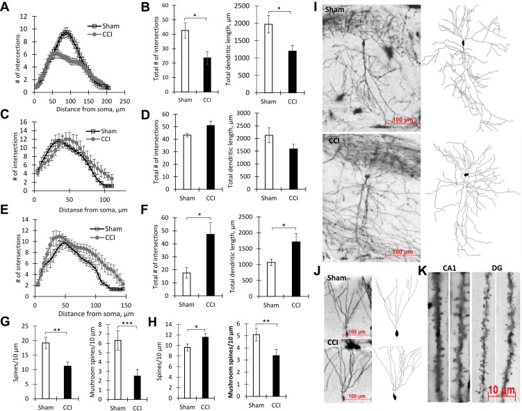
Behavioral testing revealed reduced locomotor activity as well as impaired working and long-term memory in mice with a ligated nerve. We revealed changes in the dendritic tree morphology in CA1 and the dentate gyrus hippocampal subregions. We found the atrophy of the CA1 pyramidal neurons and an increase in the dendritic tree complexity in DG. Moreover, changes in the density of dendritic spines were observed in these regions. In addition, we revealed increased expression of the Arc protein in DG granule neurons and decreased surface expression of AMPA receptors within the hippocampus. Decreased AMPA receptors expression underlies observed altered dendrite arborization and dendritic spines morphology.
We found that pain information entering the hippocampus causes neuronal plasticity changes. The changes in neurite arborization, dendritic length and dendritic spines morphology as well as protein expression are observed within the hippocampal regions involved in the processing of pain information. Moreover, changes in the dendrite morphology in hippocampal subregions are different due to the anatomical and functional heterogeneity of the hippocampus. Apparently, the detected morphological and biochemical changes can underlie the observed hippocampus-dependent behavioral and cognitive impairment in animals with neuropathic pain.
神经性疼痛表现为多种感觉症状以及高级神经活动障碍的组合,如记忆缺陷、焦虑、抑郁、快感缺失等。这表明包括海马体在内的脑结构参与了神经性疼痛的发病机制。阐明神经性疼痛认知和情感症状背后的中枢敏化机制,可能有助于开发针对这些常见疾病的新型有效治疗方法。本研究旨在阐明慢性神经性疼痛对认知功能及海马体潜在神经元可塑性的影响。
采用小鼠右后肢坐骨神经慢性缩窄损伤作为神经性疼痛模型。通过热痛觉过敏和机械性痛觉过敏确认神经性疼痛的存在。使用高尔基-考克斯染色法研究CA1锥体神经元和齿状回颗粒神经元的形态。通过免疫组织化学和酶联免疫吸附测定法测定海马体蛋白浓度。
行为测试显示,神经结扎小鼠的运动活动减少,工作记忆和长期记忆受损。我们发现CA1区和齿状回海马亚区的树突形态发生了变化。我们发现CA1锥体神经元萎缩,齿状回树突的复杂性增加。此外,在这些区域观察到树突棘密度的变化。此外,我们发现齿状回颗粒神经元中Arc蛋白的表达增加,海马体内AMPA受体的表面表达减少。AMPA受体表达降低是观察到的树突分支和树突棘形态改变的基础。
我们发现进入海马体的疼痛信息会导致神经元可塑性变化。在参与疼痛信息处理的海马区域内观察到神经突分支、树突长度和树突棘形态以及蛋白表达的变化。此外,由于海马体的解剖和功能异质性,海马亚区的树突形态变化有所不同。显然,检测到的形态和生化变化可能是神经性疼痛动物中观察到的海马体依赖性行为和认知障碍的基础。