• 文献检索
  • 文档翻译
  • 深度研究
  • 学术资讯
  • Suppr Zotero 插件Zotero 插件
  • 邀请有礼
  • 套餐&价格
  • 历史记录
应用&插件
Suppr Zotero 插件Zotero 插件浏览器插件Mac 客户端Windows 客户端微信小程序
定价
高级版会员购买积分包购买API积分包
服务
文献检索文档翻译深度研究API 文档MCP 服务
关于我们
关于 Suppr公司介绍联系我们用户协议隐私条款
关注我们

Suppr 超能文献

核心技术专利:CN118964589B侵权必究
粤ICP备2023148730 号-1Suppr @ 2026

文献检索

告别复杂PubMed语法,用中文像聊天一样搜索,搜遍4000万医学文献。AI智能推荐,让科研检索更轻松。

立即免费搜索

文件翻译

保留排版,准确专业,支持PDF/Word/PPT等文件格式,支持 12+语言互译。

免费翻译文档

深度研究

AI帮你快速写综述,25分钟生成高质量综述,智能提取关键信息,辅助科研写作。

立即免费体验

基于锥形束计算机断层扫描的沙特亚人群下颌第一磨牙中磨牙根的患病率。

The prevalence of radix molaris in the mandibular first molars of a Saudi subpopulation based on cone-beam computed tomography.

作者信息

Al-Alawi Hassan, Al-Nazhan Saad, Al-Maflehi Nassr, Aldosimani Mazen A, Zahid Mohammed Nabil, Shihabi Ghadeer N

机构信息

Dental Department, Ministry of Health Endodontist, Huraymala General Hospital, Riyadh, Saudi Arabia.

Department of Restorative Dentistry-Endodontics, College of Dentistry, Riyadh Elm University, Riyadh, Saudi Arabia.

出版信息

Restor Dent Endod. 2019 Nov 14;45(1):e1. doi: 10.5395/rde.2020.45.e1. eCollection 2020 Feb.

DOI:10.5395/rde.2020.45.e1
PMID:32110531
原文链接:https://pmc.ncbi.nlm.nih.gov/articles/PMC7030963/
Abstract

OBJECTIVES

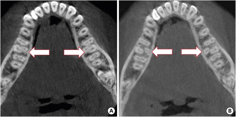
The purpose of this study was to determine the incidence of radix molaris (RM) (entomolaris and paramolaris) in the mandibular first permanent molars of a sample Saudi Arabian subpopulation using cone-beam computed tomography (CBCT).

MATERIALS AND METHODS

A total of 884 CBCT images of 427 male and 457 female Saudi citizens (age 16 to 70 years) were collected from the radiology department archives of 4 dental centers. A total of 450 CBCT images of 741 mature mandibular first molars that met the inclusion criteria were reviewed. The images were viewed at high resolution by 3 examiners and were analyzed with Planmeca Romexis software (version 5.2).

RESULTS

Thirty-three (4.5%) mandibular first permanent molars had RM, mostly on the distal side. The incidence of radix entomolaris (EM) was 4.3%, while that of radix paramolaris was 0.3%. The RM roots had one canal and occurred more unilaterally. No significant difference in root configuration was found between males and females ( > 0.05). Types I and III EM root canal configurations were most common, while type B was the only RP configuration observed.

CONCLUSIONS

The incidence of RM in the mandibular first molars of this Saudi subpopulation was 4.5%. Identification of the supernumerary root can avoid missing the canal associated with the root during root canal treatment.

摘要

目的

本研究的目的是使用锥形束计算机断层扫描(CBCT)确定沙特阿拉伯一个亚人群样本的下颌第一恒磨牙中磨牙根(RM)(远中磨牙根和副磨牙根)的发生率。

材料与方法

从4个牙科中心的放射科档案中收集了427名男性和457名女性沙特公民(年龄16至70岁)的884张CBCT图像。对符合纳入标准的741颗成熟下颌第一磨牙的450张CBCT图像进行了回顾。由3名检查人员以高分辨率查看图像,并使用Planmeca Romexis软件(版本5.2)进行分析。

结果

33颗(4.5%)下颌第一恒磨牙有RM,大多位于远中侧。远中磨牙根(EM)的发生率为4.3%,而副磨牙根的发生率为0.3%。RM根有一个根管,且多为单侧出现。男性和女性在牙根形态上未发现显著差异(>0.05)。I型和III型EM根管形态最为常见,而B型是观察到的唯一RP形态。

结论

该沙特亚人群下颌第一磨牙中RM的发生率为4.5%。识别额外牙根可避免在根管治疗过程中遗漏与该牙根相关的根管。

https://cdn.ncbi.nlm.nih.gov/pmc/blobs/7aa9/7030963/dab68819d096/rde-45-e1-g002.jpg
https://cdn.ncbi.nlm.nih.gov/pmc/blobs/7aa9/7030963/3d51fb2f32a5/rde-45-e1-g001.jpg
https://cdn.ncbi.nlm.nih.gov/pmc/blobs/7aa9/7030963/dab68819d096/rde-45-e1-g002.jpg
https://cdn.ncbi.nlm.nih.gov/pmc/blobs/7aa9/7030963/3d51fb2f32a5/rde-45-e1-g001.jpg
https://cdn.ncbi.nlm.nih.gov/pmc/blobs/7aa9/7030963/dab68819d096/rde-45-e1-g002.jpg

相似文献

1
The prevalence of radix molaris in the mandibular first molars of a Saudi subpopulation based on cone-beam computed tomography.基于锥形束计算机断层扫描的沙特亚人群下颌第一磨牙中磨牙根的患病率。
Restor Dent Endod. 2019 Nov 14;45(1):e1. doi: 10.5395/rde.2020.45.e1. eCollection 2020 Feb.
2
Radix molaris is a hidden truth of mandibular first permanent molars: A descriptive- analytic study using cone beam computed tomography.下颌第一恒磨牙根分叉区隐匿形态的研究:锥形束 CT 分析
Technol Health Care. 2023;31(5):1957-1968. doi: 10.3233/THC-230300.
3
Prevalence and Classification of C-Shaped Canal and Radix in Mandibular Molars Using Cone-Beam Computed Tomography on Mexican Population.使用锥形束计算机断层扫描对墨西哥人群下颌磨牙C形根管和牙根的患病率及分类研究
Dent J (Basel). 2024 Jul 10;12(7):212. doi: 10.3390/dj12070212.
4
Evaluation of the Frequency and Anatomy of Radix Entomolaris and Paramolaris in Lower Molars by Cone Beam Computed Tomography (CBCT) in Northern Iran, 2020-2021: A Retrospective Study.2020 - 2021年伊朗北部锥形束计算机断层扫描(CBCT)评估下颌磨牙远中根及额外根的频率和解剖结构:一项回顾性研究
Cureus. 2023 Oct 11;15(10):e46854. doi: 10.7759/cureus.46854. eCollection 2023 Oct.
5
Evaluation of radix entomolaris in mandibular first and second molars using cone-beam computed tomography and review of the literature.利用锥形束 CT 评价下颌第一、二磨牙的齿根位置:文献复习
Oral Radiol. 2020 Oct;36(4):320-326. doi: 10.1007/s11282-019-00406-0. Epub 2019 Aug 21.
6
Prevalence of Radix Entomolaris in Mandibular Permanent Molars Analyzed by Cone-Beam CT in the Saudi Population of Ha'il Province.采用锥形束CT分析沙特哈伊勒省人群下颌恒磨牙中磨牙根的患病率。
Cureus. 2023 Oct 14;15(10):e47034. doi: 10.7759/cureus.47034. eCollection 2023 Oct.
7
Root and root canal morphology of permanent mandibular first and second molars in a Kuwaiti population: A retrospective cone-beam computed tomography study.科威特人群中恒下颌第一和第二磨牙的牙根及根管形态:一项回顾性锥形束计算机断层扫描研究
Saudi Dent J. 2023 May;35(4):345-353. doi: 10.1016/j.sdentj.2023.03.008. Epub 2023 Mar 24.
8
Root canal morphology of permanent teeth in a Malaysian subpopulation using cone-beam computed tomography.马来西亚人群恒牙根尖形态的锥形束 CT 研究。
BMC Oral Health. 2019 Jan 14;19(1):14. doi: 10.1186/s12903-019-0710-z.
9
Cone-beam computed tomography as advanced diagnostic aid in endodontic treatment of molars with multiple canals: Two case reports.锥形束计算机断层扫描作为多根管磨牙牙髓治疗的先进诊断辅助手段:两例病例报告。
J Conserv Dent. 2017 Jul-Aug;20(4):273-277. doi: 10.4103/0972-0707.219194.
10
A Cone Beam Computed Tomography-Based Investigation of the Frequency and Pattern of Radix Entomolaris in the Saudi Arabian Population.基于锥形束计算机断层扫描的沙特阿拉伯人群根尖木叶状突频率和形态的研究。
Medicina (Kaunas). 2023 Nov 17;59(11):2025. doi: 10.3390/medicina59112025.

引用本文的文献

1
Radix Entomolaris and Complex Incisor Anatomy in a Saudi Cohort: A Retrospective Study.沙特人群中磨牙根与复杂切牙解剖结构:一项回顾性研究
Diagnostics (Basel). 2025 Jul 6;15(13):1721. doi: 10.3390/diagnostics15131721.
2
Prevalence of radix molaris in mandibular molars of a subpopulation of Brazil's Northeast region: a cross-sectional CBCT study.巴西东北部地区一个亚群体下颌磨牙中第三磨牙牙根的患病率:一项横断面CBCT研究
Sci Rep. 2025 Jul 2;15(1):22651. doi: 10.1038/s41598-025-06790-4.
3
Evaluation of the variations of mandibular molars and the distance from root apex to the inferior alveolar nerve in Saudi Sub-population: Three-dimensional radiographic evaluation.

本文引用的文献

1
CBCT study of mandibular first molars with a distolingual root in Koreans.韩国人下颌第一磨牙远中舌侧根的锥形束计算机断层扫描研究
Restor Dent Endod. 2018 Jul 30;43(3):e33. doi: 10.5395/rde.2018.43.e33. eCollection 2018 Aug.
2
Prevalence of Three Rooted Permanent Mandibular First Molars in Haryana (North Indian) Population.哈里亚纳邦(印度北部)人群中三根恒下颌第一磨牙的患病率
Contemp Clin Dent. 2017 Jan-Mar;8(1):38-41. doi: 10.4103/ccd.ccd_699_16.
3
Prevalence of Extra Roots in Permanent Mandibular First Molars in Iranian Population: A CBCT Analysis.
沙特亚人群下颌磨牙变异及根尖至下牙槽神经距离的评估:三维影像学评估
PLoS One. 2025 Feb 19;20(2):e0317053. doi: 10.1371/journal.pone.0317053. eCollection 2025.
4
Prevalence of in a northern Peruvian population evaluated by cone-beam tomography.通过锥形束断层扫描评估的秘鲁北部人群中的患病率。 (你提供的原文“Prevalence of in a northern Peruvian population evaluated by cone-beam tomography.”中“Prevalence of ”后面缺少具体内容,请确认原文是否准确完整。)
Heliyon. 2024 Aug 8;10(16):e35919. doi: 10.1016/j.heliyon.2024.e35919. eCollection 2024 Aug 30.
5
Comprehensive Analysis of Mandibular First Molar Root and Canal Morphology in Saudi Patients Using Cone Beam Computed Tomography (CBCT).使用锥形束计算机断层扫描(CBCT)对沙特患者下颌第一磨牙的牙根和根管形态进行综合分析。
Med Sci Monit. 2024 Aug 8;30:e945364. doi: 10.12659/MSM.945364.
6
Assessment of the root and canal morphology in the permanent dentition of Saudi Arabian population using cone beam computed and micro-computed tomography - a systematic review.使用锥形束计算机断层扫描和微计算机断层扫描评估沙特阿拉伯人群恒牙的根管和根管形态:系统评价。
BMC Oral Health. 2024 Mar 16;24(1):343. doi: 10.1186/s12903-024-04101-3.
7
A Cone Beam Computed Tomography-Based Investigation of the Frequency and Pattern of Radix Entomolaris in the Saudi Arabian Population.基于锥形束计算机断层扫描的沙特阿拉伯人群根尖木叶状突频率和形态的研究。
Medicina (Kaunas). 2023 Nov 17;59(11):2025. doi: 10.3390/medicina59112025.
8
Evaluation of the Frequency and Anatomy of Radix Entomolaris and Paramolaris in Lower Molars by Cone Beam Computed Tomography (CBCT) in Northern Iran, 2020-2021: A Retrospective Study.2020 - 2021年伊朗北部锥形束计算机断层扫描(CBCT)评估下颌磨牙远中根及额外根的频率和解剖结构:一项回顾性研究
Cureus. 2023 Oct 11;15(10):e46854. doi: 10.7759/cureus.46854. eCollection 2023 Oct.
9
Prevalence of Radix Entomolaris in Mandibular Permanent Molars Analyzed by Cone-Beam CT in the Saudi Population of Ha'il Province.采用锥形束CT分析沙特哈伊勒省人群下颌恒磨牙中磨牙根的患病率。
Cureus. 2023 Oct 14;15(10):e47034. doi: 10.7759/cureus.47034. eCollection 2023 Oct.
10
Root and root canal morphology of permanent mandibular first and second molars in a Kuwaiti population: A retrospective cone-beam computed tomography study.科威特人群中恒下颌第一和第二磨牙的牙根及根管形态:一项回顾性锥形束计算机断层扫描研究
Saudi Dent J. 2023 May;35(4):345-353. doi: 10.1016/j.sdentj.2023.03.008. Epub 2023 Mar 24.
伊朗人群恒牙下颌第一磨牙额外根的患病率:一项CBCT分析。
Iran Endod J. 2017 Winter;12(1):70-73. doi: 10.22037/iej.2017.14.
4
Prevalence and morphometric analysis of three-rooted mandibular first molars in a Brazilian subpopulation.巴西亚人群中三根下颌第一磨牙的患病率及形态计量分析。
J Appl Oral Sci. 2016 Sep-Oct;24(5):535-542. doi: 10.1590/1678-775720150511.
5
Incidence of Radix Entomolaris in Mandibular First Molars in Palestinian Population: A Clinical Investigation.巴勒斯坦人群下颌第一磨牙远中根发生率的临床研究。
Int Sch Res Notices. 2014 Nov 24;2014:405601. doi: 10.1155/2014/405601. eCollection 2014.
6
Evaluation of the effect of tooth type and canal configuration on crown size in mandibular premolars by cone-beam computed tomography.通过锥形束计算机断层扫描评估下颌前磨牙的牙齿类型和根管形态对冠部大小的影响。
Iran Endod J. 2013 Fall;8(4):153-6. Epub 2013 Oct 7.
7
Use of cone-beam computed tomography to evaluate root and canal morphology of mandibular first and second molars in Turkish individuals.应用锥形束 CT 评估土耳其人群下颌第一、二磨牙的根管及牙根形态。
Med Oral Patol Oral Cir Bucal. 2013 Jul 1;18(4):e737-44. doi: 10.4317/medoral.18473.
8
Morphological changes in the crown of mandibular molars with an additional distolingual root.下颌磨牙额外远中舌根的牙冠形态变化。
Arch Oral Biol. 2013 Mar;58(3):248-53. doi: 10.1016/j.archoralbio.2012.07.015. Epub 2012 Sep 13.
9
Use of cone-beam computed tomography to evaluate root and canal morphology of mandibular molars in Chinese individuals.应用锥形束 CT 评估中国人下颌磨牙的根管及牙根形态。
Int Endod J. 2011 Nov;44(11):990-9. doi: 10.1111/j.1365-2591.2011.01904.x. Epub 2011 Jun 10.
10
Evaluation of the root and canal morphology of mandibular first permanent molars in a western Chinese population by cone-beam computed tomography.应用锥形束 CT 评估中国西部人群下颌第一恒磨牙的根管及牙根形态。
J Endod. 2010 Nov;36(11):1786-9. doi: 10.1016/j.joen.2010.08.016. Epub 2010 Sep 17.