• 文献检索
  • 文档翻译
  • 深度研究
  • 学术资讯
  • Suppr Zotero 插件Zotero 插件
  • 邀请有礼
  • 套餐&价格
  • 历史记录
应用&插件
Suppr Zotero 插件Zotero 插件浏览器插件Mac 客户端Windows 客户端微信小程序
定价
高级版会员购买积分包购买API积分包
服务
文献检索文档翻译深度研究API 文档MCP 服务
关于我们
关于 Suppr公司介绍联系我们用户协议隐私条款
关注我们

Suppr 超能文献

核心技术专利:CN118964589B侵权必究
粤ICP备2023148730 号-1Suppr @ 2026

文献检索

告别复杂PubMed语法,用中文像聊天一样搜索,搜遍4000万医学文献。AI智能推荐,让科研检索更轻松。

立即免费搜索

文件翻译

保留排版,准确专业,支持PDF/Word/PPT等文件格式,支持 12+语言互译。

免费翻译文档

深度研究

AI帮你快速写综述,25分钟生成高质量综述,智能提取关键信息,辅助科研写作。

立即免费体验

恒牙下颌第一磨牙的峡部、副根管及牙根弯曲方向:一项计算机断层扫描研究。

Isthmuses, accessory canals, and the direction of root curvature in permanent mandibular first molars: an computed tomography study.

作者信息

Chuppani Dastgerdi Aria, Navabi Manizheh, Rakhshan Vahid

机构信息

Department of Dental Morphology, Dental Branch, Islamic Azad University, Tehran, Iran.

Department of Removable Prosthodontics, Dental Faculty, Tehran Medical Sciences, Islamic Azad University, Tehran, Iran.

出版信息

Restor Dent Endod. 2019 Dec 12;45(1):e7. doi: 10.5395/rde.2020.45.e7. eCollection 2020 Feb.

DOI:10.5395/rde.2020.45.e7
PMID:32110536
原文链接:https://pmc.ncbi.nlm.nih.gov/articles/PMC7030962/
Abstract

OBJECTIVES

This study was performed to assess the anatomy of mandibular first molars.

MATERIALS AND METHODS

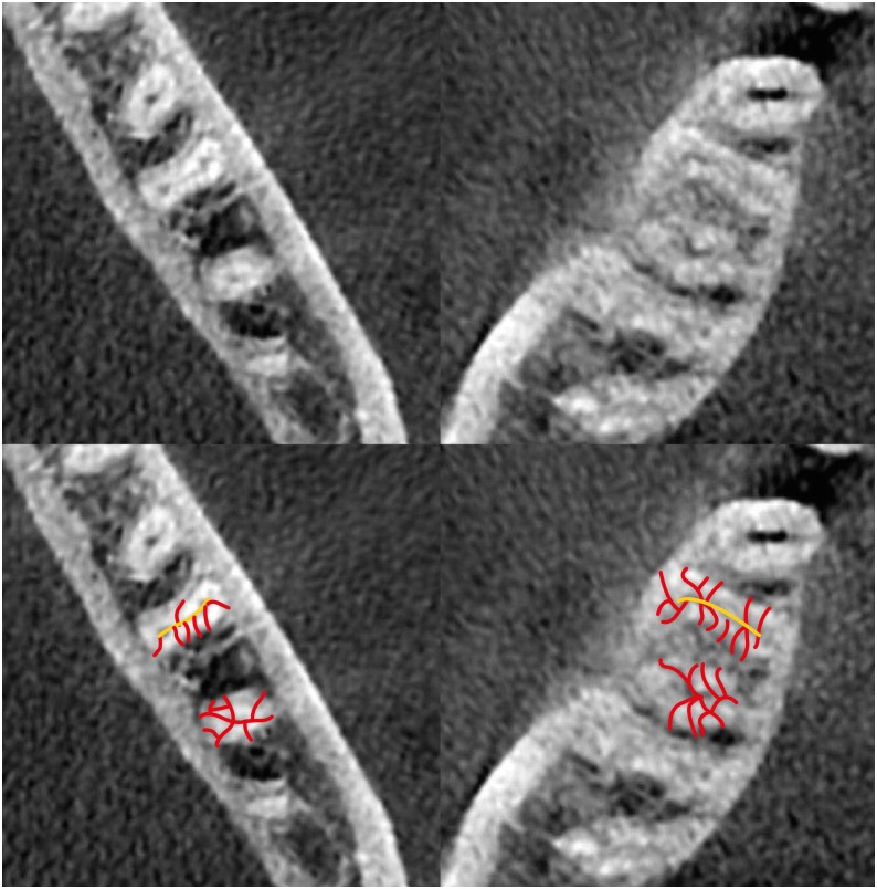
In this study, cone-beam computed tomography (CBCT) volumes of 312 bilateral intact first mandibular molars from 156 patients (79 men and 77 women; average age, 35.6 ± 11.2 years) were investigated in terms of the direction of each canal's curvature in the buccolingual and mesiodistal dimensions (direction of the position of the apex in relation to the longitudinal axis of the root), the presence of an isthmus (a narrow, ribbon-shaped communication between 2 root canals) in 3 segments (0-2, 2-4, and 4-6 mm) from the apex), and the presence and number of accessory canals (smaller canals besides the main root canals, connecting the pulp to the periodontium). Data were analyzed statistically (α = 0.05).

RESULTS

Mesiolingual canals were mostly buccally and distally inclined, while mesiobuccal and distolingual canals were mostly distally curved. Isthmuses were more common in younger patients (χ test, < 0.05). The average numbers of accessory canals in the apical, middle, and coronal segments were 9.9 ± 4.2, 6.9 ± 2.9, and 9.3 ± 3.0 canals per segment, respectively (analysis of variance, < 0.001). Age and sex were not associated with the number of accessory canals ( > 0.05).

CONCLUSIONS

The complex anatomy of these teeth deserves attention during non-surgical or surgical endodontic treatment. Around the apex, isthmuses might be more prevalent in younger and female individuals.

摘要

目的

本研究旨在评估下颌第一磨牙的解剖结构。

材料与方法

本研究中,对156例患者(79名男性和77名女性;平均年龄35.6±11.2岁)的312颗双侧完整下颌第一磨牙的锥形束计算机断层扫描(CBCT)图像进行研究,分析各根管在颊舌向和近远中向的弯曲方向(根尖相对于牙根纵轴的位置方向)、根尖上方3个节段(0 - 2、2 - 4和4 - 6 mm)峡部(两根管之间狭窄的带状连通区)的存在情况,以及副根管(除主根管外连接牙髓和牙周组织的较小根管)的存在情况和数量。数据进行统计学分析(α = 0.05)。

结果

近中舌根管大多向颊侧和远中倾斜,而近中颊根管和远中舌根管大多向远中弯曲。峡部在年轻患者中更常见(χ检验,<0.05)。根尖段、中段和冠段副根管的平均数量分别为每段9.9±4.2、6.9±2.9和9.3±3.0条(方差分析,<0.001)。年龄和性别与副根管数量无关(>0.05)。

结论

在非手术或手术牙髓治疗过程中,这些牙齿复杂的解剖结构值得关注。在根尖周围,峡部可能在年轻个体和女性中更普遍。

https://cdn.ncbi.nlm.nih.gov/pmc/blobs/e1e5/7030962/897c4dab121b/rde-45-e7-g001.jpg

相似文献

1
Isthmuses, accessory canals, and the direction of root curvature in permanent mandibular first molars: an computed tomography study.恒牙下颌第一磨牙的峡部、副根管及牙根弯曲方向:一项计算机断层扫描研究。
Restor Dent Endod. 2019 Dec 12;45(1):e7. doi: 10.5395/rde.2020.45.e7. eCollection 2020 Feb.
2
Coronal root canal morphology of permanent two-rooted mandibular first molars with novel 3D measurements.具有新型三维测量的恒双根下颌第一磨牙的冠状根管形态。
Int Endod J. 2020 Feb;53(2):167-175. doi: 10.1111/iej.13220. Epub 2019 Oct 6.
3
Prevalence and morphometric analysis of three-rooted mandibular first molars in a Brazilian subpopulation.巴西亚人群中三根下颌第一磨牙的患病率及形态计量分析。
J Appl Oral Sci. 2016 Sep-Oct;24(5):535-542. doi: 10.1590/1678-775720150511.
4
Micro-computed tomography study of the internal anatomy of mesial root canals of mandibular molars.下颌磨牙近中根管内部解剖的微计算机断层研究。
J Endod. 2011 Dec;37(12):1682-6. doi: 10.1016/j.joen.2011.08.001. Epub 2011 Sep 9.
5
Geometric Analysis of the Distolingual Root and Canal in Mandibular First Molars: A Micro-computed Tomographic Study.下颌第一磨牙远中舌根的几何形态分析:显微 CT 研究。
J Endod. 2021 May;47(5):779-786. doi: 10.1016/j.joen.2021.02.006. Epub 2021 Feb 18.
6
Micro-computed tomographic analysis of the mesial root of mandibular first molars with bifid apex.下颌第一磨牙近中颊根双根畸形的微焦点 CT 分析
Arch Oral Biol. 2020 Sep;117:104792. doi: 10.1016/j.archoralbio.2020.104792. Epub 2020 Jun 8.
7
Morphological Characteristics and Classification of Mandibular First Molars Having 2 Distal Roots or Canals: 3-Dimensional Biometric Analysis Using Cone-beam Computed Tomography in a Korean Population.下颌第一磨牙具有 2 个远中根或根管的形态特征和分类:韩国人群的锥形束 CT 三维生物测量分析。
J Endod. 2018 Jan;44(1):46-50. doi: 10.1016/j.joen.2017.08.005. Epub 2017 Oct 21.
8
Three-dimensional analysis of coronal root canal morphology of 136 permanent mandibular first molars by micro-computed tomography.利用显微计算机断层扫描对136颗恒牙下颌第一磨牙的冠部根管形态进行三维分析。
J Dent Sci. 2022 Jan;17(1):482-489. doi: 10.1016/j.jds.2021.07.021. Epub 2021 Aug 13.
9
[Cone-beam computed tomography for determination of mesial root canal curvatures of human mandibular first molars].[锥形束计算机断层扫描用于测定人类下颌第一磨牙近中根管弯曲度]
Nan Fang Yi Ke Da Xue Xue Bao. 2018 Jul 30;38(7):824-829. doi: 10.3969/j.issn.1673-4254.2018.07.09.
10
Evaluation of Distolingual Canal/Roots in Mandibular Molars and Mesiobuccal Canals in Maxillary Molars by CBCT.CBCT对下颌磨牙远中舌侧根管/牙根及上颌磨牙近中颊侧根管的评估
Open Dent J. 2017 Jun 30;11:360-366. doi: 10.2174/1874210601711010360. eCollection 2017.

引用本文的文献

1
Comprehensive analysis of root canal morphology in maxillary premolars among the Pakistani subpopulation: a CBCT-based study.基于 CBCT 的巴基斯坦人群上颌前磨牙根管形态的综合分析研究。
Eur J Med Res. 2024 Jul 27;29(1):391. doi: 10.1186/s40001-024-01990-6.
2
Exploring age and gender variations in root canal morphology of maxillary premolars in Saudi sub population: a cross-sectional CBCT study.探讨沙特亚族群上颌前磨牙根管形态的年龄和性别差异:一项横断面 CBCT 研究。
BMC Oral Health. 2024 May 9;24(1):543. doi: 10.1186/s12903-024-04310-w.
3
Micro-CT and histological examination of accessory canals in 34 equine cheek teeth.

本文引用的文献

1
Anatomy of Permanent‎ ‎Mandibular‎ First‎ Molars in a Selected Iranian Population Using ‎Cone-beam Computed Tomography.使用锥形束计算机断层扫描对选定伊朗人群下颌第一恒磨牙的解剖结构研究
Iran Endod J. 2018 Spring;13(2):251-256. doi: 10.22037/iej.v13i2.19035.
2
Accuracy of Cone-beam Computed Tomography in Comparison with Standard Method in Evaluating Root Canal Morphology: An Study.锥形束计算机断层扫描与标准方法在评估根管形态方面的准确性比较:一项研究。
Iran Endod J. 2018 Spring;13(2):181-187. doi: 10.22037/iej.v13i2.18614.
3
Root and Canal Morphology of Mandibular Molars in a Selected Iranian Population Using Cone-Beam Computed Tomography.
34颗马颊齿副根管的显微CT及组织学检查
Front Vet Sci. 2024 Apr 10;11:1396871. doi: 10.3389/fvets.2024.1396871. eCollection 2024.
4
Diagnostic value of cone beam computed tomography for root canal morphology assessment - a micro-CT based comparison.锥形束计算机断层扫描在根管形态评估中的诊断价值——基于微 CT 的比较。
Clin Oral Investig. 2024 Mar 8;28(3):201. doi: 10.1007/s00784-024-05580-y.
使用锥形束计算机断层扫描对选定伊朗人群下颌磨牙的根管形态进行研究。
Iran Endod J. 2017 Spring;12(2):143-148. doi: 10.22037/iej.2017.29.
4
Root Canal Anatomy and Morphology of Mandibular First Molars in a Selected Iranian Population: An Study.特定伊朗人群下颌第一磨牙的根管解剖与形态:一项研究
Iran Endod J. 2017 Winter;12(1):87-91. doi: 10.22037/iej.2017.18.
5
Radicular anatomy of permanent mandibular second molars in an Iranian population: A preliminary study.伊朗人群中恒牙下颌第二磨牙的根管解剖:一项初步研究。
Dent Res J (Isfahan). 2016 Jul-Aug;13(4):362-6. doi: 10.4103/1735-3327.187883.
6
Identification of Apical and Cervical Curvature Radius of Human Molars.人类磨牙根尖和牙颈曲率半径的识别
Braz Dent J. 2015 Jul-Aug;26(4):351-6. doi: 10.1590/0103-64402013x0252.
7
Evaluation of isthmus prevalence, location, and types in mesial roots of mandibular molars in the Iranian Population.伊朗人群下颌磨牙近中根峡部的发生率、位置及类型评估。
Dent Res J (Isfahan). 2014 Mar;11(2):251-6.
8
Comparison of alternative image reformatting techniques in micro-computed tomography and tooth clearing for detailed canal morphology.微计算机断层扫描和牙齿透明化中用于详细根管形态的替代图像重格式化技术的比较
J Endod. 2014 Mar;40(3):417-22. doi: 10.1016/j.joen.2013.09.014. Epub 2013 Oct 25.
9
An In Vitro Study of the Number of Distal Roots and Canals in mandibular First Molars in Iranian Population.伊朗人群下颌第一磨牙远中根及根管数量的体外研究
Iran Endod J. 2008 Winter;2(4):126-30. Epub 2008 Jan 10.
10
Use of cone-beam computed tomography to evaluate root and canal morphology of mandibular first and second molars in Turkish individuals.应用锥形束 CT 评估土耳其人群下颌第一、二磨牙的根管及牙根形态。
Med Oral Patol Oral Cir Bucal. 2013 Jul 1;18(4):e737-44. doi: 10.4317/medoral.18473.