• 文献检索
  • 文档翻译
  • 深度研究
  • 学术资讯
  • Suppr Zotero 插件Zotero 插件
  • 邀请有礼
  • 套餐&价格
  • 历史记录
应用&插件
Suppr Zotero 插件Zotero 插件浏览器插件Mac 客户端Windows 客户端微信小程序
定价
高级版会员购买积分包购买API积分包
服务
文献检索文档翻译深度研究API 文档MCP 服务
关于我们
关于 Suppr公司介绍联系我们用户协议隐私条款
关注我们

Suppr 超能文献

核心技术专利:CN118964589B侵权必究
粤ICP备2023148730 号-1Suppr @ 2026

文献检索

告别复杂PubMed语法,用中文像聊天一样搜索,搜遍4000万医学文献。AI智能推荐,让科研检索更轻松。

立即免费搜索

文件翻译

保留排版,准确专业,支持PDF/Word/PPT等文件格式,支持 12+语言互译。

免费翻译文档

深度研究

AI帮你快速写综述,25分钟生成高质量综述,智能提取关键信息,辅助科研写作。

立即免费体验

伊朗人群下颌磨牙近中根峡部的发生率、位置及类型评估。

Evaluation of isthmus prevalence, location, and types in mesial roots of mandibular molars in the Iranian Population.

作者信息

Mehrvarzfar Payman, Akhlagi Nahid Mohammadzade, Khodaei Fatemeh, Shojaee Golnaz, Shirazi Sara

机构信息

Department of Endodontics, Dental branch, Islamic Azad University, Tehran, Iran.

出版信息

Dent Res J (Isfahan). 2014 Mar;11(2):251-6.

PMID:24932198
原文链接:https://pmc.ncbi.nlm.nih.gov/articles/PMC4052653/
Abstract

BACKGROUND

Management of canal isthmus is considered as an important factor for successful endodontic treatment. Accordingly, this study was designed to determine the prevalence, location, and types of isthmus in mesial root canals of extracted mandibular molars in a sample of Iranian population.

MATERIALS AND METHODS

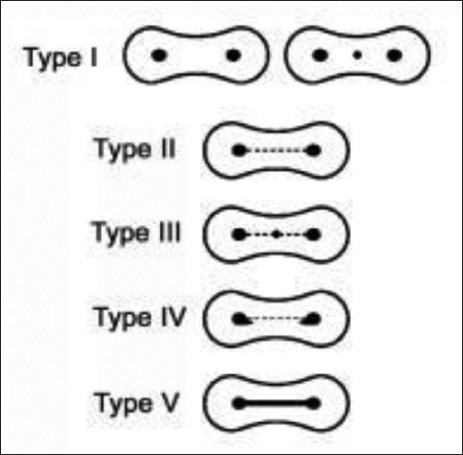
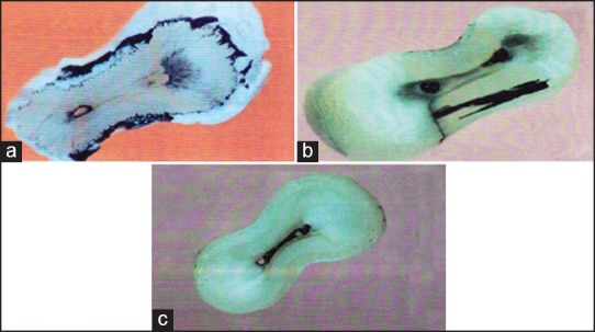
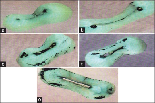
In this cross-sectional descriptive study, 60 extracted molars with two mesial canals were included. The samples were initially decoronated and then, roots were sectioned horizontally at 2, 4, and 6 mm levels from the apex via a low-speed handpiece with a thin metallic disk and finally prepared and stained with Indian ink. All sections were examined using a stereomicroscope at a magnification of ×30. Prevalence, location, and types of isthmus were evaluated based on the classifications by Kim and Teixeira and all data were statistically analyzed by the chi-squared test. The statistical significance level was established at 0.05.

RESULTS

Eighty-three percent of extracted mandibular molars had an isthmus at the mesial root. This prevalence increased with distance from the apex, that is, 92% at 6 mm from the apex and 70% at 2 mm from the apex. A statistically significant difference was found between the sections at 2 and 6 mm from the apex (P < 0.05), but no other significant differences between other levels (P > 0.05).

CONCLUSION

Isthmus is very common in the mesial roots of the mandibular permanent molars in the Iranian population, with the highest prevalence in the 6 mm distance from the root apex. Therefore, detection, cleaning, and filling of these apical 6 mm isthmuses are of great benefit in modern endodontics.

摘要

背景

根管峡部的处理被认为是牙髓治疗成功的一个重要因素。因此,本研究旨在确定伊朗人群样本中拔除的下颌磨牙近中根管峡部的发生率、位置和类型。

材料与方法

在这项横断面描述性研究中,纳入了60颗有两条近中根管的拔除磨牙。样本首先去除牙冠,然后使用带有薄金属盘的低速手机从根尖分别在2mm、4mm和6mm水平进行水平切片,最后用印度墨水进行制备和染色。所有切片均在放大30倍的体视显微镜下检查。根据Kim和Teixeira的分类评估峡部的发生率、位置和类型,所有数据均采用卡方检验进行统计学分析。统计学显著性水平设定为0.05。

结果

83%的拔除下颌磨牙近中根存在峡部。这种发生率随着与根尖距离的增加而升高,即距根尖6mm处为92%,距根尖2mm处为70%。在距根尖2mm和6mm处的切片之间发现有统计学显著差异(P < 0.05),但其他水平之间无其他显著差异(P > 0.05)。

结论

峡部在伊朗人群下颌恒磨牙近中根中非常常见,在距根尖6mm处发生率最高。因此,在现代牙髓病学中,对这些根尖6mm峡部的检测、清理和充填具有很大益处。

https://cdn.ncbi.nlm.nih.gov/pmc/blobs/6c2e/4052653/3f14ef08a813/DRJ-11-251-g006.jpg
https://cdn.ncbi.nlm.nih.gov/pmc/blobs/6c2e/4052653/da7ecf3d7b48/DRJ-11-251-g002.jpg
https://cdn.ncbi.nlm.nih.gov/pmc/blobs/6c2e/4052653/58654ffdd8bf/DRJ-11-251-g005.jpg
https://cdn.ncbi.nlm.nih.gov/pmc/blobs/6c2e/4052653/3f14ef08a813/DRJ-11-251-g006.jpg
https://cdn.ncbi.nlm.nih.gov/pmc/blobs/6c2e/4052653/da7ecf3d7b48/DRJ-11-251-g002.jpg
https://cdn.ncbi.nlm.nih.gov/pmc/blobs/6c2e/4052653/58654ffdd8bf/DRJ-11-251-g005.jpg
https://cdn.ncbi.nlm.nih.gov/pmc/blobs/6c2e/4052653/3f14ef08a813/DRJ-11-251-g006.jpg

相似文献

1
Evaluation of isthmus prevalence, location, and types in mesial roots of mandibular molars in the Iranian Population.伊朗人群下颌磨牙近中根峡部的发生率、位置及类型评估。
Dent Res J (Isfahan). 2014 Mar;11(2):251-6.
2
A preliminary in vitro study of the incidence and position of the root canal isthmus in maxillary and mandibular first molars.一项关于上颌和下颌第一磨牙根管峡部发生率及位置的初步体外研究。
Int Endod J. 2003 Apr;36(4):276-80. doi: 10.1046/j.1365-2591.2003.00638.x.
3
The isthmuses of the mesial root of mandibular molars: a micro-computed tomographic study.下颌磨牙近中根峡部:一项显微计算机断层扫描研究。
Int Endod J. 2005 Aug;38(8):558-63. doi: 10.1111/j.1365-2591.2005.00994.x.
4
Prevalence and morphological analysis of isthmuses in mandibular molars of the Indian population: A micro-computed tomographic study.印度人群下颌磨牙峡部的患病率及形态学分析:一项显微计算机断层扫描研究。
J Conserv Dent Endod. 2023 Sep-Oct;26(5):584-589. doi: 10.4103/JCDE.JCDE_52_23. Epub 2023 Sep 16.
5
[A micro-computed tomographic study of the isthmus in the root canal system of mandibular first molar].[下颌第一磨牙根管系统峡部的显微计算机断层扫描研究]
Zhonghua Kou Qiang Yi Xue Za Zhi. 2009 Jan;44(1):11-4.
6
Micro-computed tomographic analysis of the mesial root of mandibular first molars with bifid apex.下颌第一磨牙近中颊根双根畸形的微焦点 CT 分析
Arch Oral Biol. 2020 Sep;117:104792. doi: 10.1016/j.archoralbio.2020.104792. Epub 2020 Jun 8.
7
Apical anatomy in mesial and mesiobuccal roots of permanent first molars.恒牙第一磨牙近中根和近中颊根的根尖解剖结构。
J Endod. 2005 May;31(5):364-8. doi: 10.1097/01.don.0000145425.73364.91.
8
Topographic Analysis of the Isthmus in Mesiobuccal and Mesial Roots of First Molars in a South Korean Population.韩国人群第一磨牙近中颊根和近中舌根峡部的地形分析。
Sci Rep. 2020 Jan 27;10(1):1247. doi: 10.1038/s41598-020-58364-1.
9
Root Canal Anatomy and Morphology of Mandibular First Molars in a Selected Iranian Population: An Study.特定伊朗人群下颌第一磨牙的根管解剖与形态:一项研究
Iran Endod J. 2017 Winter;12(1):87-91. doi: 10.22037/iej.2017.18.
10
Root and Canal Morphology of Mandibular Molars in a Selected Iranian Population Using Cone-Beam Computed Tomography.使用锥形束计算机断层扫描对选定伊朗人群下颌磨牙的根管形态进行研究。
Iran Endod J. 2017 Spring;12(2):143-148. doi: 10.22037/iej.2017.29.

引用本文的文献

1
Micro-computed tomographic evaluation of root and canal anatomy of maxillary first premolars in Iraqi sub-population.伊拉克亚人群上颌第一前磨牙牙根及根管解剖结构的显微计算机断层扫描评估
Sci Rep. 2025 Mar 28;15(1):10821. doi: 10.1038/s41598-025-94590-1.
2
Presence and distribution of voids after using the single cone obturation technique with different sealer placement methods in canals with an isthmus.在有峡部的根管中使用不同封闭剂放置方法的单锥充填技术后空隙的存在及分布情况。
J Dent Sci. 2025 Jan;20(1):286-291. doi: 10.1016/j.jds.2024.07.014. Epub 2024 Jul 25.
3
Prevalence and morphological analysis of isthmuses in mandibular molars of the Indian population: A micro-computed tomographic study.

本文引用的文献

1
Canal and isthmus debridement efficacy of the VPro EndoSafe negative-pressure irrigation technique.VPro EndoSafe 负压冲洗技术的管腔和峡部清创效果。
J Endod. 2012 Dec;38(12):1631-4. doi: 10.1016/j.joen.2012.09.002. Epub 2012 Oct 17.
2
Canal and isthmus debridement efficacy using a sonic irrigation technique in a closed-canal system.采用超声冲洗技术对闭合根管系统的根管和峡部进行清创的疗效。
J Endod. 2012 Sep;38(9):1265-8. doi: 10.1016/j.joen.2012.05.009. Epub 2012 Jul 20.
3
One- versus two-visit endodontic treatment of teeth with apical periodontitis: a histobacteriologic study.
印度人群下颌磨牙峡部的患病率及形态学分析:一项显微计算机断层扫描研究。
J Conserv Dent Endod. 2023 Sep-Oct;26(5):584-589. doi: 10.4103/JCDE.JCDE_52_23. Epub 2023 Sep 16.
4
Root canal isthmi and interorifice distance in human permanent teeth of an Indian subpopulation using cone-beam computed tomography: A retrospective cross-sectional study.使用锥形束计算机断层扫描对印度亚人群恒牙根管峡部和根管口间距离的研究:一项回顾性横断面研究
J Conserv Dent. 2020 Nov-Dec;23(6):563-570. doi: 10.4103/JCD.JCD_576_20. Epub 2021 Feb 11.
5
[Cone-beam computed tomography for determination of mesial root canal curvatures of human mandibular first molars].[锥形束计算机断层扫描用于测定人类下颌第一磨牙近中根管弯曲度]
Nan Fang Yi Ke Da Xue Xue Bao. 2018 Jul 30;38(7):824-829. doi: 10.3969/j.issn.1673-4254.2018.07.09.
6
Isthmuses, accessory canals, and the direction of root curvature in permanent mandibular first molars: an computed tomography study.恒牙下颌第一磨牙的峡部、副根管及牙根弯曲方向:一项计算机断层扫描研究。
Restor Dent Endod. 2019 Dec 12;45(1):e7. doi: 10.5395/rde.2020.45.e7. eCollection 2020 Feb.
7
Isthmus Incidence in Human Permanent Mandibular First Molars of a South Indian Population: A Cone-Beam Computerized Tomographic Study.南印度人群恒牙下颌第一磨牙峡部发生率:一项锥形束计算机断层扫描研究
J Pharm Bioallied Sci. 2019 May;11(Suppl 2):S468-S473. doi: 10.4103/JPBS.JPBS_80_19.
8
Evaluation of the Prevalence of Complete Isthmii in Permanent Teeth Using Cone-Beam Computed Tomography.使用锥形束计算机断层扫描评估恒牙中完全峡部的患病率。
Iran Endod J. 2017 Fall;12(4):426-431. doi: 10.22037/iej.v12i4.17175.
一次性与两次性根管治疗术治疗根尖周炎患牙的疗效比较:一项组织细菌学研究。
J Endod. 2012 Aug;38(8):1040-52. doi: 10.1016/j.joen.2012.04.010. Epub 2012 Jun 12.
4
A high-resolution computed tomographic study of changes in root canal isthmus area by instrumentation and root filling.根管器械预备和根管充填后根管峡部面积变化的高分辨率 CT 研究。
J Endod. 2011 Feb;37(2):223-7. doi: 10.1016/j.joen.2010.10.012.
5
Apical surgery: endoscopic findings at the resection level of 168 consecutively treated roots.根尖手术:168 例连续治疗根的切除水平内镜检查结果。
Int Endod J. 2011 Apr;44(4):290-302. doi: 10.1111/j.1365-2591.2010.01811.x. Epub 2011 Jan 12.
6
Three-dimensional morphologic analysis of isthmuses in the mesial roots of mandibular molars.下颌磨牙近中根峡部的三维形态分析。
J Endod. 2010 Nov;36(11):1866-9. doi: 10.1016/j.joen.2010.08.030. Epub 2010 Sep 17.
7
Canal and isthmus debridement efficacies of two irrigant agitation techniques in a closed system.两种闭系统冲洗液搅拌技术的管腔和峡部清创效果。
Int Endod J. 2010 Dec;43(12):1077-90. doi: 10.1111/j.1365-2591.2010.01778.x. Epub 2010 Aug 19.
8
Root and canal morphology of mandibular second molars in an Indian population.下颌第二磨牙的根管和根形态在印度人群中的研究。
J Endod. 2010 Aug;36(8):1319-22. doi: 10.1016/j.joen.2010.04.001. Epub 2010 May 23.
9
Laser-activated irrigation within root canals: cleaning efficacy and flow visualization.激光激活根管内冲洗:清洗效果和流动可视化。
Int Endod J. 2009 Dec;42(12):1077-83. doi: 10.1111/j.1365-2591.2009.01634.x.
10
Ultrastructural examination of failed molar retreatment with secondary apical periodontitis: an examination of endodontic biofilms in an endodontic retreatment failure.继发根尖周炎的磨牙再治疗失败的超微结构检查:对根管再治疗失败中根管生物膜的检查
J Endod. 2009 Sep;35(9):1303-9. doi: 10.1016/j.joen.2009.05.035.