Department of Nuclear Medicine, University Hospital Essen, University Duisburg-Essen, Hufelandstr. 55, 45147, Essen, Germany.
High-Field and Hybrid MR Imaging, University Hospital Essen, University Duisburg-Essen, Hufelandstr. 55, 45147, Essen, Germany.
Eur J Nucl Med Mol Imaging. 2020 Sep;47(10):2269-2279. doi: 10.1007/s00259-020-04738-6. Epub 2020 Mar 3.
This study evaluates the quantitative effect of improved MR-based attenuation correction (AC), including bone segmentation and the HUGE method for truncation correction in PET/MR whole-body hybrid imaging specifically of oncologic patients with bone metastasis and using various radiotracers.
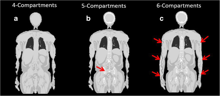
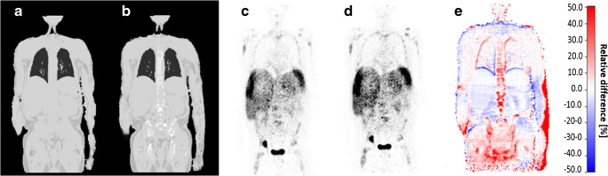
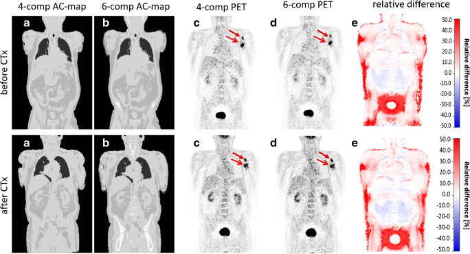
Twenty-three patients that underwent altogether 28 whole-body PET/MR examinations with findings of bone metastasis were included in this study. Different radiotracers (F-FDG, Ga-PSMA, Ga-DOTATOC, I-MIBG) were injected according to appropriate clinical indications. Each of the 28 whole-body PET datasets was reconstructed three times using AC with (1) standard four-compartment μ-maps (background air, lung, muscle, and soft tissue), (2) five-compartment μ-maps (adding bone), and (3) six-compartment μ-maps (adding bone and HUGE truncation correction). The SUV of each detected bone lesion was measured in each reconstruction to evaluate the quantitative impact of improved MR-based AC. Relative difference images between four- and six-compartment μ-maps were calculated. MR-based HUGE truncation correction was compared with the PET-based MLAA truncation correction method in all patients.
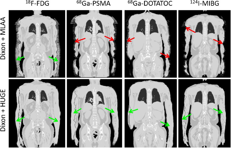
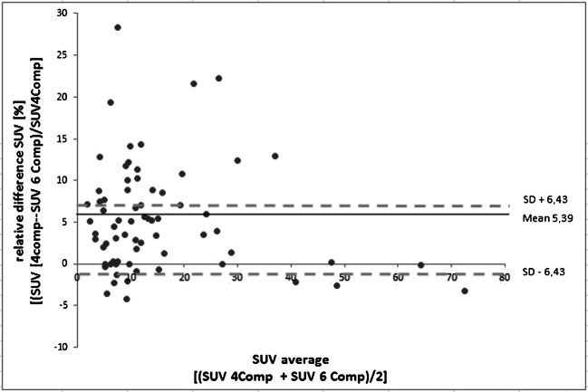
Overall, 69 bone lesions were detected and evaluated. The mean increase in relative difference over all 69 lesions in SUV was 5.4 ± 6.4% when comparing the improved six-compartment AC with the standard four-compartment AC. Maximal relative difference of 28.4% was measured in one lesion. Truncation correction with HUGE worked robust and resulted in realistic body contouring in all 28 exams and for all 4 different radiotracers. Truncation correction with MLAA revealed overestimations of arm tissue volume in all PET/MR exams with F-FDG radiotracer and failed in all other exams with radiotracers Ga-PSMA, Ga-DOTATOC, and I- MIBG due to limitations in body contour detection.
Improved MR-based AC, including bone segmentation and HUGE truncation correction in whole-body PET/MR on patients with bone lesions and using various radiotracers, is important to ensure best possible diagnostic image quality and accurate PET quantification. The HUGE method for truncation correction based on MR worked robust and results in realistic body contouring, independent of the radiotracers used.
本研究评估了改进的基于磁共振(MR)的衰减校正(AC)的定量效果,包括骨分割和 HUGE 方法用于截断校正,特别是在使用各种放射性示踪剂的骨转移肿瘤患者的全身 PET/MR 混合成像中。
本研究纳入了 23 例共 28 例全身 PET/MR 检查的患者,这些患者均发现有骨转移。根据适当的临床指征,分别注射不同的放射性示踪剂(F-FDG、Ga-PSMA、Ga-DOTATOC、I-MIBG)。28 例全身 PET 数据集的每个数据集均使用以下 3 种 AC 方法进行重建:(1)标准的四室 μ 图谱(背景空气、肺、肌肉和软组织);(2)五室 μ 图谱(增加骨);(3)六室 μ 图谱(增加骨和 HUGE 截断校正)。在每种重建中测量每个检测到的骨病变的 SUV,以评估改进的基于 MR 的 AC 的定量影响。计算四室和六室 μ 图谱之间的相对差异图像。在所有患者中,将基于 MR 的 HUGE 截断校正与基于 PET 的 MLAA 截断校正方法进行比较。
总体上,共检测到并评估了 69 个骨病变。当将改进的六室 AC 与标准的四室 AC 进行比较时,69 个病变的 SUV 中相对差异的平均增加为 5.4%±6.4%。在一个病变中测量到最大的相对差异为 28.4%。HUGE 截断校正方法在所有 28 例检查和所有 4 种不同放射性示踪剂中均工作可靠,并产生了逼真的身体轮廓。使用 F-FDG 放射性示踪剂的所有 PET/MR 检查中,MLAA 截断校正均导致手臂组织体积的高估,而在使用 Ga-PSMA、Ga-DOTATOC 和 I-MIBG 放射性示踪剂的其他所有检查中,由于身体轮廓检测的限制,MLAA 截断校正均失败。
在有骨病变的患者中使用各种放射性示踪剂进行全身 PET/MR 检查时,改进的基于 MR 的 AC,包括骨分割和 HUGE 截断校正,对于确保最佳的诊断图像质量和准确的 PET 定量非常重要。基于 MR 的 HUGE 截断校正方法工作可靠,可产生逼真的身体轮廓,与使用的放射性示踪剂无关。