Lagziel Tomer, Sylvester Scott, Hultman Charles S, Asif Mohammed
Plastic Surgery, The Johns Hopkins University School of Medicine, Baltimore, USA.
Cureus. 2020 Jan 22;12(1):e6736. doi: 10.7759/cureus.6736.
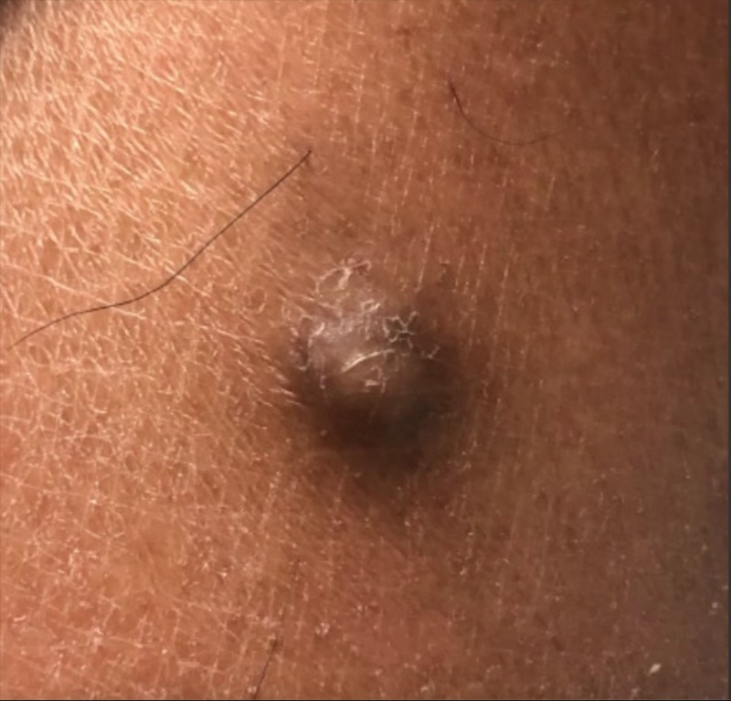
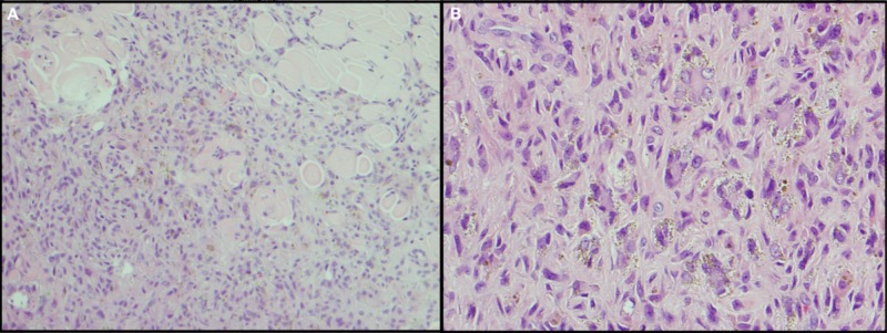
A dermatofibroma (DF) is a common, benign tumor composed of fibroblastic and histiocytic cells. DF presents clinically with several different reported variants. One rare variant is hemosiderotic DF (HDF), which is made up of small blood vessels and hemosiderin deposits. HDF can be indistinguishable, clinically, from melanoma, making the use of other pathological tools crucial in the diagnosis. We report the case of a 25-year-old male medical student from the Caribbean who presented to our clinic with a single asymptomatic pigmented cystic lesion on his left posterior calf. The cystic lesion was excised surgically. Histopathology examination of the excised mass revealed a moderately cellular, poorly demarcated, dermal, fibrohistiocytic proliferation. Pathology consultation confirmed a diagnosis of HDF.
皮肤纤维瘤(DF)是一种常见的良性肿瘤,由成纤维细胞和组织细胞组成。DF在临床上有几种不同的报道变体。一种罕见的变体是含铁血黄素性皮肤纤维瘤(HDF),它由小血管和含铁血黄素沉积组成。HDF在临床上可能与黑色素瘤难以区分,这使得使用其他病理工具对诊断至关重要。我们报告了一名来自加勒比地区的25岁男性医学生的病例,他因左小腿后侧出现一个无症状的色素性囊性病变前来我们诊所就诊。该囊性病变通过手术切除。对切除肿块的组织病理学检查显示为中度细胞性、边界不清的真皮纤维组织细胞增生。病理会诊确诊为HDF。