Department of Gastroenterology, SMG-SNU Boramae Medical Center, Seoul National University of College of Medicine, Seoul, South Korea.
Department of Gastroenterology, Gangneung Asan Hospital, University of Ulsan College of Medicine, Gangneung, South Korea.
BMC Gastroenterol. 2020 Mar 5;20(1):51. doi: 10.1186/s12876-020-01197-z.
Lymph node (LN) metastasis is negligible in early gastric cancer (EGC) within expanded criteria for endoscopic submucosal dissection (ESD). However, regional lymph nodes in abdominal CT scans are sometimes enlarged in patients with EGC within the expanded criteria for endoscopic submucosal dissection (ESD). In this study, we investigated the clinical significance of regional lymph node enlargement on abdominal CT scan in patients with EGC within the expanded criteria for ESD.
From December 2010 to April 2015, among 301 patients with EGC within the ESD expanded criteria, 47 patients with regional lymph node enlargement shown by abdominal CT scan were prospectively enrolled. We performed surgical resection or periodic follow-up with abdominal CT scans and upper endoscopy every 6 months to evaluate whether the enlarged lymph nodes are due to metastasis or a reactive change.
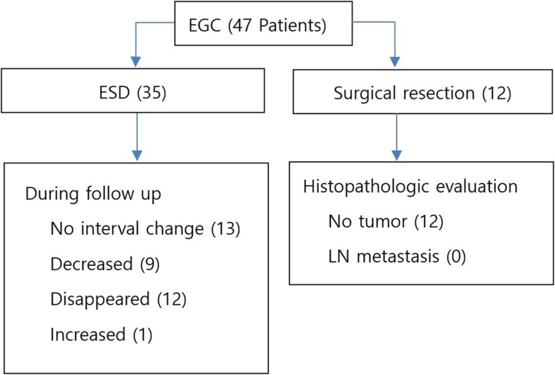
The mean age of the 47 patients (38 males, 9 female) was 64.8 years. The enlarged lymph nodes were usually single (26/47, 44.6%) and sized as follows: 11 nodes were ≤ 5 mm, 19 were 6-10 mm, and 17 were ≥ 10 mm. Four of the 47 patients initially underwent surgical resection, and 8 patients underwent surgical resection after ESD. However, there was no lymph node metastasis in surgical specimens. Thirty-five patients received ESD and periodically followed up at a median duration of 56 months (IQR: 44-59 month). The enlarged lymph node disappeared in 12 of 35 patients, decreased in 9 patients and remained the same size in 13 patients, and increased in 1 patient.
Regional lymph node enlargement on abdominal CT scan in patients within expanded criteria for ESD of ECG may be not due to metastasis but a reactive change.
在扩大内镜黏膜下剥离术(ESD)适应证的早期胃癌(EGC)中,淋巴结(LN)转移可忽略不计。然而,在扩大内镜黏膜下剥离术适应证的 EGC 患者中,腹部 CT 扫描有时会显示区域淋巴结肿大。在本研究中,我们探讨了在扩大内镜黏膜下剥离术适应证的 EGC 患者中,腹部 CT 扫描显示区域淋巴结肿大的临床意义。
2010 年 12 月至 2015 年 4 月,在 301 例扩大内镜黏膜下剥离术适应证的 EGC 患者中,前瞻性纳入 47 例腹部 CT 扫描显示区域淋巴结肿大的患者。我们对这些患者进行了手术切除或定期随访,每 6 个月进行一次腹部 CT 扫描和上消化道内镜检查,以评估肿大的淋巴结是否为转移或反应性改变。
47 例患者(男 38 例,女 9 例)的平均年龄为 64.8 岁。肿大的淋巴结通常为单个(26/47,44.6%),大小如下:11 个淋巴结≤5mm,19 个淋巴结为 6-10mm,17 个淋巴结≥10mm。47 例患者中有 4 例最初接受了手术切除,8 例患者在 ESD 后接受了手术切除。然而,手术标本中未见淋巴结转移。35 例患者接受了 ESD 治疗,并在中位随访 56 个月(IQR:44-59 个月)后定期随访。35 例患者中,12 例肿大的淋巴结消失,9 例淋巴结缩小,13 例淋巴结大小保持不变,1 例淋巴结增大。
在扩大内镜黏膜下剥离术适应证的 EGC 患者中,腹部 CT 扫描显示区域淋巴结肿大可能不是转移,而是反应性改变。