• 文献检索
  • 文档翻译
  • 深度研究
  • 学术资讯
  • Suppr Zotero 插件Zotero 插件
  • 邀请有礼
  • 套餐&价格
  • 历史记录
应用&插件
Suppr Zotero 插件Zotero 插件浏览器插件Mac 客户端Windows 客户端微信小程序
定价
高级版会员购买积分包购买API积分包
服务
文献检索文档翻译深度研究API 文档MCP 服务
关于我们
关于 Suppr公司介绍联系我们用户协议隐私条款
关注我们

Suppr 超能文献

核心技术专利:CN118964589B侵权必究
粤ICP备2023148730 号-1Suppr @ 2026

文献检索

告别复杂PubMed语法,用中文像聊天一样搜索,搜遍4000万医学文献。AI智能推荐,让科研检索更轻松。

立即免费搜索

文件翻译

保留排版,准确专业,支持PDF/Word/PPT等文件格式,支持 12+语言互译。

免费翻译文档

深度研究

AI帮你快速写综述,25分钟生成高质量综述,智能提取关键信息,辅助科研写作。

立即免费体验

近视眼中黄斑厚度与视盘旁萎缩的相关性。

Association of macular thickness with parapapillary atrophy in myopic eyes.

机构信息

Department of Ophthalmology and Research Institute of Medical Sciences, Chonnam National University Medical School and Hospital, 42 Jebong-ro, Dong-gu, Gwangju, 61469, South Korea.

Department of Ophthalmology, Yanbian University Hospital, Yanji, Jilin, China.

出版信息

BMC Ophthalmol. 2020 Mar 6;20(1):93. doi: 10.1186/s12886-020-01362-8.

DOI:10.1186/s12886-020-01362-8
PMID:32143676
原文链接:https://pmc.ncbi.nlm.nih.gov/articles/PMC7060613/
Abstract

BACKGROUND

To investigate whether macular structure could be affected by axial elongation and to determine the association between macular intraretinal thickness and the microstructure of β-zone parapapillary atrophy (PPA) in myopic eyes.

METHODS

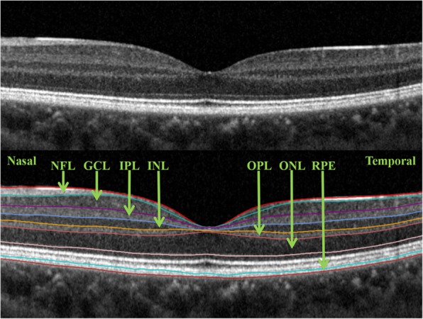
The study recruited 113 healthy myopic subjects (113 eyes). Images of the macula, subfoveal choroid, and optic nerve head were acquired using spectral-domain optical coherence tomography (SD-OCT). An automatic segmentation algorithm was used to segment the macular images into 7 intraretinal layers. PPA widths with and without Bruch's membrane (PPA and PPA, respectively) were evaluated. Linear regression analysis was performed to evaluate the association between macular intraretinal thickness and axial length and the microstructure of PPA.

RESULTS

An increase in axial length was associated with a decrease in whole macular thickness of the peripheral region and an increase in whole macular thickness of the central region. Thickness alterations of the macular intraretinal layers were most apparent in the peripheral region. A significant correlation was found between PPA width and macular intraretinal layer thickness, whereas no significant correlation was found between PPA width and macular intraretinal layer thickness. Moreover, both PPA and PPA widths significantly correlated with subfoveal choroidal thickness.

CONCLUSIONS

Macular intraretinal layer thickness may be affected by PPA width. These findings indicate that the microstructure of PPA should be considered when evaluating the macula in patient with myopia and glaucoma.

摘要

背景

探讨轴向伸长是否会影响黄斑结构,并确定近视眼中黄斑视网膜内厚度与β区神经纤维层旁视网膜萎缩(PPA)微观结构之间的关系。

方法

本研究招募了 113 名健康近视受试者(113 只眼)。使用频域光学相干断层扫描(SD-OCT)获取黄斑、中心凹下脉络膜和视盘的图像。采用自动分割算法将黄斑图像分割为 7 个视网膜内部分层。评估有无布鲁赫膜(PPA 和 PPA)的 PPA 宽度。采用线性回归分析评估黄斑视网膜内厚度与眼轴长度以及 PPA 微观结构之间的关系。

结果

眼轴长度的增加与周边区域全黄斑厚度的减少和中心区域全黄斑厚度的增加相关。黄斑视网膜内各层的厚度变化在周边区域最为明显。PPA 宽度与黄斑视网膜内各层厚度之间存在显著相关性,而 PPA 宽度与黄斑视网膜内各层厚度之间无显著相关性。此外,PPA 和 PPA 宽度均与中心凹下脉络膜厚度显著相关。

结论

黄斑视网膜内各层厚度可能受 PPA 宽度的影响。这些发现表明,在评估近视和青光眼患者的黄斑时,应考虑 PPA 的微观结构。

https://cdn.ncbi.nlm.nih.gov/pmc/blobs/cb5b/7060613/f09b88337495/12886_2020_1362_Fig3_HTML.jpg
https://cdn.ncbi.nlm.nih.gov/pmc/blobs/cb5b/7060613/23295aab0a2f/12886_2020_1362_Fig1_HTML.jpg
https://cdn.ncbi.nlm.nih.gov/pmc/blobs/cb5b/7060613/f5dc45cd4ca6/12886_2020_1362_Fig2_HTML.jpg
https://cdn.ncbi.nlm.nih.gov/pmc/blobs/cb5b/7060613/f09b88337495/12886_2020_1362_Fig3_HTML.jpg
https://cdn.ncbi.nlm.nih.gov/pmc/blobs/cb5b/7060613/23295aab0a2f/12886_2020_1362_Fig1_HTML.jpg
https://cdn.ncbi.nlm.nih.gov/pmc/blobs/cb5b/7060613/f5dc45cd4ca6/12886_2020_1362_Fig2_HTML.jpg
https://cdn.ncbi.nlm.nih.gov/pmc/blobs/cb5b/7060613/f09b88337495/12886_2020_1362_Fig3_HTML.jpg

相似文献

1
Association of macular thickness with parapapillary atrophy in myopic eyes.近视眼中黄斑厚度与视盘旁萎缩的相关性。
BMC Ophthalmol. 2020 Mar 6;20(1):93. doi: 10.1186/s12886-020-01362-8.
2
Microstructure of Parapapillary Atrophy Is Associated With Parapapillary Microvasculature in Myopic Eyes.近视眼中的神经旁萎缩的微观结构与神经旁微血管有关。
Am J Ophthalmol. 2018 Aug;192:157-168. doi: 10.1016/j.ajo.2018.05.022. Epub 2018 May 30.
3
Microstructure of β-zone parapapillary atrophy and rate of retinal nerve fiber layer thinning in primary open-angle glaucoma.原发性开角型青光眼β-区视盘旁萎缩的微观结构与视网膜神经纤维层变薄的关系。
Ophthalmology. 2014 Jul;121(7):1341-9. doi: 10.1016/j.ophtha.2014.01.008. Epub 2014 Feb 22.
4
Parapapillary atrophy and changes in the optic nerve head and posterior pole in high myopia.高度近视的视盘旁萎缩及视神经头和后极部改变。
Sci Rep. 2020 Mar 12;10(1):4607. doi: 10.1038/s41598-020-61485-2.
5
En Face Optical Coherence Tomography Imaging of Beta and Gamma Parapapillary Atrophy in High Myopia.高度近视β和γ 视盘旁萎缩的 En Face 光学相干断层扫描成像。
Ophthalmol Glaucoma. 2019 Jan-Feb;2(1):55-62. doi: 10.1016/j.ogla.2018.11.008. Epub 2018 Nov 30.
6
Topographic Correlation Between Juxtapapillary Choroidal Thickness and Microstructure of Parapapillary Atrophy.黄斑旁脉络膜厚度与旁中心凹萎缩结构的眼底地形图相关性。
Ophthalmology. 2016 Sep;123(9):1965-73. doi: 10.1016/j.ophtha.2016.05.006. Epub 2016 Jun 16.
7
Factors associated with deep circulation in the peripapillary chorioretinal atrophy zone in normal-tension glaucoma with myopic disc.与正常眼压性近视盘青光眼伴周边脉络膜视网膜萎缩区深部循环相关的因素。
Acta Ophthalmol. 2018 May;96(3):e290-e297. doi: 10.1111/aos.13621. Epub 2017 Nov 24.
8
Spectral-domain optical coherence tomography of β-zone peripapillary atrophy: influence of myopia and glaucoma.β 区视盘旁萎缩的光谱域光相干断层扫描:近视和青光眼的影响。
Invest Ophthalmol Vis Sci. 2012 Mar 21;53(3):1499-505. doi: 10.1167/iovs.11-8572.
9
Longitudinal Changes of Optic Nerve Head and Peripapillary Structure during Childhood Myopia Progression on OCT: Boramae Myopia Cohort Study Report 1.基于 OCT 的儿童近视进展过程中视盘和视盘周围结构的纵向变化:Boramae 近视队列研究报告 1。
Ophthalmology. 2018 Aug;125(8):1215-1223. doi: 10.1016/j.ophtha.2018.01.026. Epub 2018 Mar 14.
10
Macular Thickness Profiles of Intraretinal Layers in Myopia Evaluated by Ultrahigh-Resolution Optical Coherence Tomography.通过超高分辨率光学相干断层扫描评估近视患者视网膜内层的黄斑厚度分布
Am J Ophthalmol. 2015 Jul;160(1):53-61.e2. doi: 10.1016/j.ajo.2015.03.012. Epub 2015 Mar 20.

引用本文的文献

1
Changes in choriocapillaris blood flow density and associated factors affecting axial elongation in Chinese children and adolescents with mild to moderate myopia: a cross-sectional study.中国轻中度近视儿童及青少年脉络膜毛细血管血流密度变化及影响眼轴长度的相关因素:一项横断面研究
BMC Ophthalmol. 2025 Jul 28;25(1):426. doi: 10.1186/s12886-025-04272-9.

本文引用的文献

1
Structure-function Relationship in Advanced Glaucoma After Reaching the RNFL Floor.高级青光眼达到神经纤维层底部后的结构-功能关系。
J Glaucoma. 2019 Nov;28(11):1006-1011. doi: 10.1097/IJG.0000000000001374.
2
Normative Data for Retinal-Layer Thickness Maps Generated by Spectral-Domain OCT in a White Population.白种人群中光谱域光学相干断层扫描生成的视网膜层厚度图的规范数据。
Ophthalmol Retina. 2018 Aug;2(8):808-815.e1. doi: 10.1016/j.oret.2017.12.012. Epub 2018 Feb 6.
3
Change of β-Zone Parapapillary Atrophy During Axial Elongation: Boramae Myopia Cohort Study Report 3.
β 区视盘旁萎缩在眼轴增长过程中的变化:Boramae 近视队列研究报告 3。
Invest Ophthalmol Vis Sci. 2018 Aug 1;59(10):4020-4030. doi: 10.1167/iovs.18-24775.
4
Parapapillary Beta Zone and Gamma Zone in a Healthy Population: The Beijing Eye Study 2011.健康人群的视盘旁β区和γ区:2011 年北京眼研究。
Invest Ophthalmol Vis Sci. 2018 Jul 2;59(8):3320-3329. doi: 10.1167/iovs.18-24141.
5
Association Between Optic Nerve Head Deformation and Retinal Microvasculature in High Myopia.高度近视患者视神经头变形与视网膜微血管的关系。
Am J Ophthalmol. 2018 Apr;188:81-90. doi: 10.1016/j.ajo.2018.01.033. Epub 2018 Feb 6.
6
Optic Nerve Tilt, Crescent, Ovality, and Torsion in a Multi-Ethnic Cohort of Young Adults With and Without Myopia.有近视和无近视的多民族青年成人队列中的视神经倾斜、半月形、椭圆度和扭转
Invest Ophthalmol Vis Sci. 2017 Jun 1;58(7):3158-3171. doi: 10.1167/iovs.16-20860.
7
PARAPAPILLARY GAMMA AND DELTA ZONES IN HIGH MYOPIA.高度近视的周边部 γ 和 δ 区。
Retina. 2018 May;38(5):931-938. doi: 10.1097/IAE.0000000000001650.
8
Measurements of the parapapillary atrophy zones in en face optical coherence tomography images.在正面光学相干断层扫描图像中对视乳头周围萎缩区的测量。
PLoS One. 2017 Apr 17;12(4):e0175347. doi: 10.1371/journal.pone.0175347. eCollection 2017.
9
Is Choroidal or Scleral Thickness Related to Myopic Macular Degeneration?脉络膜或巩膜厚度与近视性黄斑变性有关吗?
Invest Ophthalmol Vis Sci. 2017 Feb 1;58(2):907-913. doi: 10.1167/iovs.16-20742.
10
Evidence-Based Treatment of Diabetic Macular Edema.糖尿病性黄斑水肿的循证治疗
Semin Ophthalmol. 2017;32(1):56-66. doi: 10.1080/08820538.2016.1228388.