Hong Wenjun, Zhao Zhiyong, Wang Dongmei, Li Ming, Tang Chaozheng, Li Zheng, Xu Rong, Chan Chetwyn C H
Department of Rehabilitation Medicine, Nanjing Drum Tower Hospital, The Affiliated Hospital of Nanjing University Medical School, Nanjing, 210008, China.
Key Laboratory for Biomedical Engineering of Ministry of Education, College of Biomedical Engineering & Instrument Science, Zhejiang University, Hangzhou, China.
Neuroimage Clin. 2020;26:102224. doi: 10.1016/j.nicl.2020.102224. Epub 2020 Feb 20.
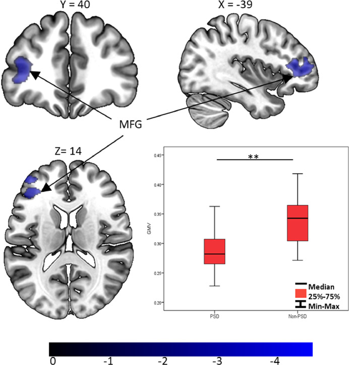
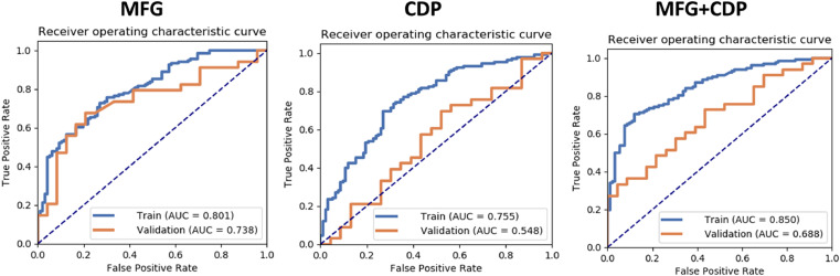
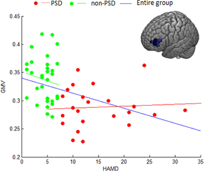
Stroke survivors are known to suffer from post-stroke depression (PSD). However, the likelihood of structural changes in the brains of PSD patients has not been explored. This study aims to extract changes in the gray matter of these patients and test how these changes account for the PSD symptoms. High-resolution T1 weighted images were collected from 23 PSD patients diagnosed with subcortical stroke. Voxel-based morphometry and support vector machine analyses were used to analyze the data. The results were compared with those collected from 33 non-PSD patients. PSD group showed decreased gray matter volume (GMV) in the left middle frontal gyrus (MFG) when compared to the non-PSD patients. Together with the clinical and demographic variables, the MFG's GMV predictive model was able to distinguish PSD from the non-PSD patients (0•70 sensitivity and 0•88 specificity). The changes in the left inferior frontal gyrus (61%) and dorsolateral prefrontal cortex (39%) suggest that the somatic/affective symptoms in PSD is likely to be due to patients' problems with understanding and appraising negative emotional stimuli. The impact brought by the reduced prefrontal to limbic system connectivity needs further exploration. These findings indicate possible systemic involvement of the frontolimbic network resulting in PSD after brain lesions which is likely to be independent from the location of the lesion. The results inform specific clinical interventions to be provided for treating depressive symptoms in post-stroke patients.
已知中风幸存者会患中风后抑郁症(PSD)。然而,尚未探究PSD患者大脑结构变化的可能性。本研究旨在提取这些患者灰质的变化,并测试这些变化如何解释PSD症状。从23名被诊断为皮质下中风的PSD患者收集了高分辨率T1加权图像。使用基于体素的形态计量学和支持向量机分析来分析数据。将结果与从33名非PSD患者收集的结果进行比较。与非PSD患者相比,PSD组左侧额中回(MFG)的灰质体积(GMV)减少。结合临床和人口统计学变量,MFG的GMV预测模型能够区分PSD患者和非PSD患者(敏感性为0.70,特异性为0.88)。左侧额下回(61%)和背外侧前额叶皮质(39%)的变化表明,PSD中的躯体/情感症状可能是由于患者在理解和评估负面情绪刺激方面存在问题。前额叶与边缘系统连接减少所带来的影响需要进一步探索。这些发现表明,脑损伤后额边缘网络可能存在系统性受累,导致PSD,这可能与病变位置无关。这些结果为治疗中风后患者的抑郁症状提供了具体的临床干预措施。