Yu Siyi, Shen Zhifu, Lai Rui, Feng Fen, Guo Baojun, Wang Zhengyan, Yang Jie, Hu Youping, Gong Liang
Department of Acupuncture & Tuina, Chengdu University of Traditional Chinese Medicine, Chengdu, China.
Department of Anesthesiology, People's Hospital of Deyang, Deyang, China.
Front Psychiatry. 2018 Dec 4;9:651. doi: 10.3389/fpsyt.2018.00651. eCollection 2018.
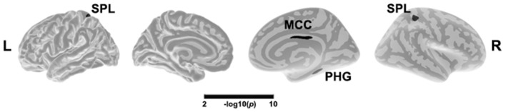
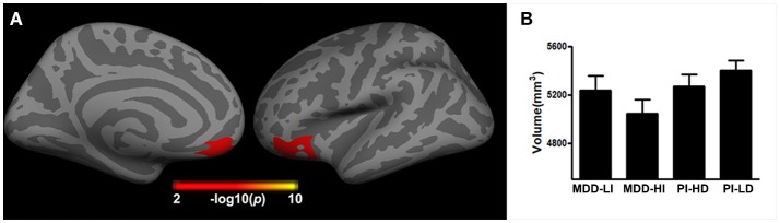
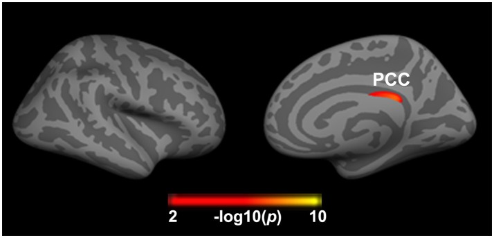
Insomnia and depression are highly comorbid symptoms in both primary insomnia (PI) and major depressive disorder (MDD). In the current study, we aimed at exploring both the homogeneous and heterogeneous brain structure alteration in PI and MDD patients. Sixty-five MDD patients and 67 matched PI patients were recruited and underwent a structural MRI scan. The subjects were sub-divided into four groups, namely MDD patients with higher or lower insomnia, and PI patients with higher or lower severe depression. A general linear model was employed to explore the changes in cortical thickness and volume as a result of depression or insomnia, and their interaction. In addition, partial correlation analysis was conducted to detect the clinical significance of the altered brain structural regions. A main effect of depression on cortical thickness was seen in the superior parietal lobe, middle cingulate cortex, and parahippocampal gyrus, while a main effect of insomnia on cortical thickness was found in the posterior cingulate cortex. Importantly, the interaction between depression and insomnia was associated with decreased gray matter volume in the right orbitofrontal cortex, i.e., patients with co-occurring depression and insomnia showed smaller brain volume in the right orbitofrontal cortex when compared to patients with lower insomnia/depression. These findings highlighted the role of the orbitofrontal cortex in the neuropathology of the comorbidity of insomnia and depression. Our findings provide new insights into the understanding of the brain mechanism underlying comorbidity of insomnia and depression.
失眠和抑郁是原发性失眠(PI)和重度抑郁症(MDD)中高度共病的症状。在本研究中,我们旨在探究PI和MDD患者大脑结构改变的同质性和异质性。招募了65名MDD患者和67名匹配的PI患者,并对其进行了结构磁共振成像扫描。受试者被分为四组,即失眠程度较高或较低的MDD患者,以及重度抑郁程度较高或较低的PI患者。采用一般线性模型来探究抑郁或失眠导致的皮质厚度和体积变化及其相互作用。此外,进行偏相关分析以检测大脑结构改变区域的临床意义。抑郁对皮质厚度的主要影响见于顶上叶、扣带中部皮质和海马旁回,而失眠对皮质厚度的主要影响见于扣带后回。重要的是,抑郁和失眠之间的相互作用与右侧眶额皮质灰质体积减少有关,即与失眠/抑郁程度较低的患者相比,同时患有抑郁和失眠的患者右侧眶额皮质的脑体积较小。这些发现突出了眶额皮质在失眠和抑郁共病神经病理学中的作用。我们的发现为理解失眠和抑郁共病的脑机制提供了新的见解。