Zhao ZhanLin, Zhang JiaYing, Liang Hong, Zhang SiYi, Shao ChunYi, Fan XianQun, Fu Yao
Department of Ophthalmology, Ninth People's Hospital, Shanghai JiaoTong University School of Medicine, Shanghai, China.
Shanghai Key Laboratory of Orbital Diseases and Ocular Oncology, Shanghai, China.
J Ophthalmol. 2020 Feb 22;2020:1349072. doi: 10.1155/2020/1349072. eCollection 2020.
To evaluate changes in corneal sensitivity and subbasal nerve density after pterygium excision.
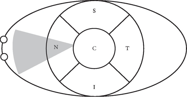
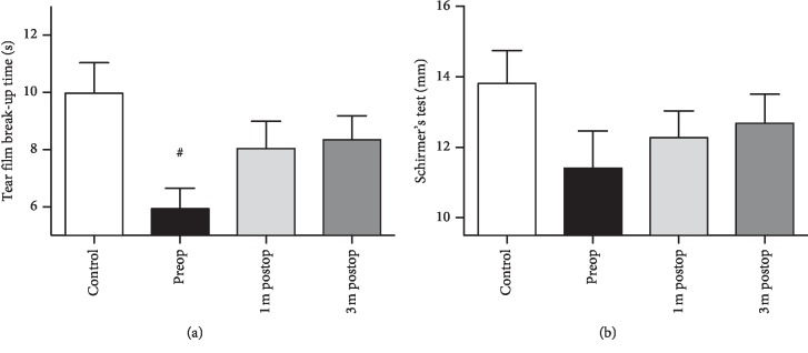
This prospective trial included 22 eyes with nasal primary pterygium and 18 controls. Corneal sensitivity was evaluated using a Cochet-Bonnet esthesiometer in the nasal, superior, temporal, inferior, and center quadrants of the cornea before surgery and 10 days, 1 month, and 3months after surgery. The central cornea was analyzed using confocal microscopy (IVCM) before surgery and 1 and 3 months after surgery. Subbasal nerve density and other nerve parameters were analyzed using NeuronJ. Nerve tortuosity was evaluated and graded in individual IVCM scans. The tear film break-up time (TBUT) test and Schirmer's test were performed before surgery, as well as 1 and 3 months after surgery. All the same tests were performed in the controls.
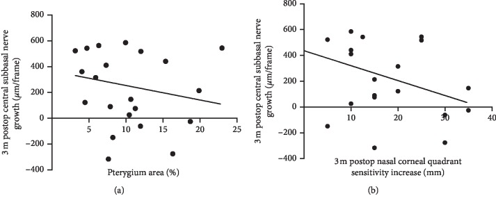
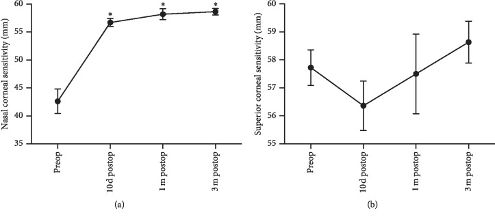
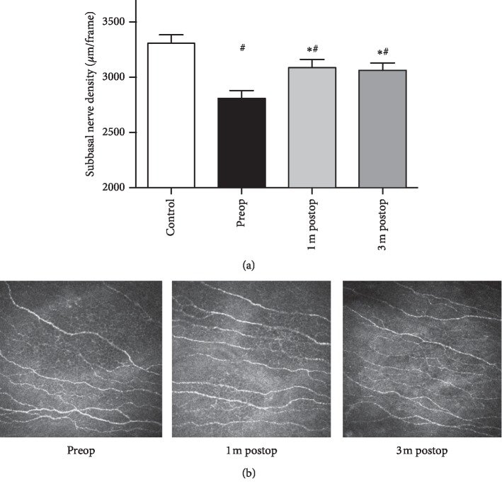
All affected eyes showed a significant increase in corneal sensitivity in the nasal corneal quadrant after surgery when compared with preoperative data ( = 37.3; < 0.01). Compared with controls, pterygium patients demonstrated decreased corneal subbasal nerve density ( < 0.01). Compared with controls, pterygium patients demonstrated decreased corneal subbasal nerve density ( < 0.01). Compared with controls, pterygium patients demonstrated decreased corneal subbasal nerve density ( < 0.01). Compared with controls, pterygium patients demonstrated decreased corneal subbasal nerve density ( = 37.3; < 0.01). Compared with controls, pterygium patients demonstrated decreased corneal subbasal nerve density ( < 0.01). Compared with controls, pterygium patients demonstrated decreased corneal subbasal nerve density ( = 37.3; < 0.01). Compared with controls, pterygium patients demonstrated decreased corneal subbasal nerve density (.
Pterygium patients demonstrated deteriorated corneal subbasal nerve fibers when compared with healthy controls in terms of nerve length, nerve trunks, and nerve branches. Therefore, pterygium excision improves corneal sensitivity and increases corneal subbasal nerve density.
评估翼状胬肉切除术后角膜敏感性和基底膜下神经密度的变化。
这项前瞻性试验纳入了22只患有鼻侧原发性翼状胬肉的眼睛和18只对照眼。在手术前以及手术后10天、1个月和3个月,使用Cochet-Bonnet触觉计评估角膜鼻侧、上方、颞侧、下方和中央象限的角膜敏感性。在手术前以及手术后1个月和3个月,使用共焦显微镜(IVCM)分析中央角膜。使用NeuronJ分析基底膜下神经密度和其他神经参数。在个体IVCM扫描中评估并分级神经迂曲度。在手术前以及手术后1个月和3个月进行泪膜破裂时间(TBUT)试验和泪液分泌试验。在对照眼中进行所有相同的测试。
与术前数据相比,所有患眼术后鼻侧角膜象限的角膜敏感性均显著增加(= 37.3;<0.01)。与对照组相比,翼状胬肉患者的角膜基底膜下神经密度降低(<0.01)。与对照组相比,翼状胬肉患者的角膜基底膜下神经密度降低(<0.01)。与对照组相比,翼状胬肉患者的角膜基底膜下神经密度降低(<0.01)。与对照组相比,翼状胬肉患者的角膜基底膜下神经密度降低(= 37.3;<0.01)。与对照组相比,翼状胬肉患者的角膜基底膜下神经密度降低(<0.01)。与对照组相比,翼状胬肉患者的角膜基底膜下神经密度降低(= 37.3;<0.01)。与对照组相比,翼状胬肉患者的角膜基底膜下神经密度降低(。
与健康对照组相比,翼状胬肉患者在神经长度、神经干和神经分支方面的角膜基底膜下神经纤维受损。因此,翼状胬肉切除术可提高角膜敏感性并增加角膜基底膜下神经密度。