• 文献检索
  • 文档翻译
  • 深度研究
  • 学术资讯
  • Suppr Zotero 插件Zotero 插件
  • 邀请有礼
  • 套餐&价格
  • 历史记录
应用&插件
Suppr Zotero 插件Zotero 插件浏览器插件Mac 客户端Windows 客户端微信小程序
定价
高级版会员购买积分包购买API积分包
服务
文献检索文档翻译深度研究API 文档MCP 服务
关于我们
关于 Suppr公司介绍联系我们用户协议隐私条款
关注我们

Suppr 超能文献

核心技术专利:CN118964589B侵权必究
粤ICP备2023148730 号-1Suppr @ 2026

文献检索

告别复杂PubMed语法,用中文像聊天一样搜索,搜遍4000万医学文献。AI智能推荐,让科研检索更轻松。

立即免费搜索

文件翻译

保留排版,准确专业,支持PDF/Word/PPT等文件格式,支持 12+语言互译。

免费翻译文档

深度研究

AI帮你快速写综述,25分钟生成高质量综述,智能提取关键信息,辅助科研写作。

立即免费体验

2 型糖尿病的临床生物标志物与光相干断层扫描血管造影参数的相关性。

Association Between Clinical Biomarkers and Optical Coherence Tomography Angiography Parameters in Type 2 Diabetes Mellitus.

机构信息

,.

出版信息

Invest Ophthalmol Vis Sci. 2020 Mar 9;61(3):4. doi: 10.1167/iovs.61.3.4.

DOI:10.1167/iovs.61.3.4
PMID:32150245
原文链接:https://pmc.ncbi.nlm.nih.gov/articles/PMC7401845/
Abstract

PURPOSE

To investigate the clinical significance of the changes in the macular microvasculature in patients with diabetes mellitus type 2 without diabetic retinopathy.

METHODS

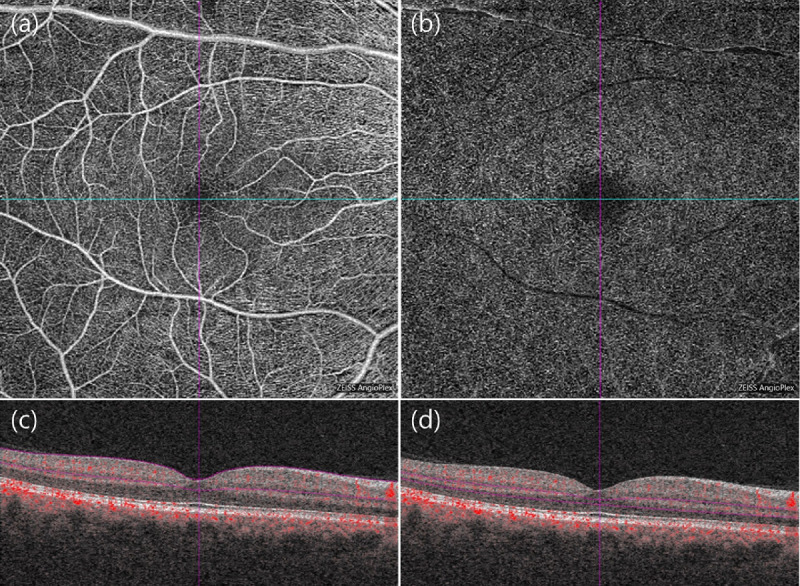
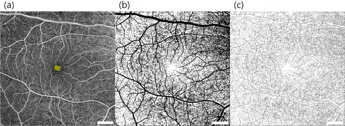
Fifty-five patients with diabetes mellitus type 2 without diabetic retinopathy and 48 healthy individuals were enrolled in a prospective cross-sectional study. We identified the changes of optical coherence tomography angiography parameters (foveal avascular zone [FAZ] area and circularity, vessel density, and perfusion index) of the 6 × 6-mm macular scan. Correlation and multiple regression analyses were performed between optical coherence tomography angiography parameters and previously known diabetes mellitus type 2-related demographic and systemic characteristics, and serum biochemical markers.

RESULTS

FAZ parameters and perfusion index of the superficial and deep vascular plexus showed significant correlation with serum insulin level, and homeostasis model assessment indices. In multiple linear regression analysis, low insulin levels predicted increased FAZ areas in both the superficial (β = -0.007; P = 0.030) and deep layers (β = -0.010; P = 0.018) and a decreased perfusion index in the deep layer (β = 0.003; P = 0.001).

CONCLUSIONS

The expansion and loss of circularity of the FAZ and the decrease in the perfusion index may be affected by insulin resistance and secretory capacity in patients with diabetes mellitus type 2 with no diabetic retinopathy.

摘要

目的

探讨 2 型糖尿病无糖尿病视网膜病变患者黄斑微血管变化的临床意义。

方法

前瞻性横断面研究纳入了 55 例 2 型糖尿病无糖尿病视网膜病变患者和 48 名健康个体。我们确定了 6×6mm 黄斑扫描的光学相干断层血管造影参数(中心凹无血管区[FAZ]面积和圆度、血管密度和灌注指数)的变化。对光学相干断层血管造影参数与先前已知的 2 型糖尿病相关的人口统计学和全身特征以及血清生化标志物进行了相关性和多元回归分析。

结果

FAZ 参数和浅层及深层血管丛的灌注指数与血清胰岛素水平和稳态模型评估指数呈显著相关。在多元线性回归分析中,低胰岛素水平预测浅层(β=-0.007;P=0.030)和深层(β=-0.010;P=0.018)FAZ 面积增大,深层灌注指数降低(β=0.003;P=0.001)。

结论

在无糖尿病视网膜病变的 2 型糖尿病患者中,FAZ 的扩大和圆度丧失以及灌注指数降低可能受胰岛素抵抗和分泌能力的影响。

https://cdn.ncbi.nlm.nih.gov/pmc/blobs/edd3/7401845/c0e98e64d9ef/iovs-61-3-4-f002.jpg
https://cdn.ncbi.nlm.nih.gov/pmc/blobs/edd3/7401845/6fa35aed6ffb/iovs-61-3-4-f001.jpg
https://cdn.ncbi.nlm.nih.gov/pmc/blobs/edd3/7401845/c0e98e64d9ef/iovs-61-3-4-f002.jpg
https://cdn.ncbi.nlm.nih.gov/pmc/blobs/edd3/7401845/6fa35aed6ffb/iovs-61-3-4-f001.jpg
https://cdn.ncbi.nlm.nih.gov/pmc/blobs/edd3/7401845/c0e98e64d9ef/iovs-61-3-4-f002.jpg

相似文献

1
Association Between Clinical Biomarkers and Optical Coherence Tomography Angiography Parameters in Type 2 Diabetes Mellitus.2 型糖尿病的临床生物标志物与光相干断层扫描血管造影参数的相关性。
Invest Ophthalmol Vis Sci. 2020 Mar 9;61(3):4. doi: 10.1167/iovs.61.3.4.
2
Optical coherence tomography angiography analysis of foveal microvascular changes and inner retinal layer thinning in patients with diabetes.糖尿病患者黄斑区微血管改变及内层视网膜变薄的光相干断层扫描血管造影分析。
Br J Ophthalmol. 2018 Sep;102(9):1226-1231. doi: 10.1136/bjophthalmol-2017-311149. Epub 2017 Dec 19.
3
Quantitative optical coherence tomography angiography of macular vascular structure and foveal avascular zone in glaucoma.青光眼黄斑血管结构及中心凹无血管区的定量光学相干断层扫描血管造影
PLoS One. 2017 Sep 21;12(9):e0184948. doi: 10.1371/journal.pone.0184948. eCollection 2017.
4
QUANTIFICATION OF RETINAL VESSEL TORTUOSITY IN DIABETIC RETINOPATHY USING OPTICAL COHERENCE TOMOGRAPHY ANGIOGRAPHY.应用光学相干断层扫描血管造影术定量分析糖尿病性视网膜病变中的视网膜血管迂曲。
Retina. 2018 May;38(5):976-985. doi: 10.1097/IAE.0000000000001618.
5
Macular vessels density in diabetic retinopathy: quantitative assessment using optical coherence tomography angiography.糖尿病视网膜病变中的黄斑血管密度:使用光学相干断层扫描血管造影术进行定量评估。
Int Ophthalmol. 2019 Aug;39(8):1845-1859. doi: 10.1007/s10792-018-1013-0. Epub 2018 Sep 7.
6
Determinants of Quantitative Optical Coherence Tomography Angiography Metrics in Patients with Diabetes.糖尿病患者定量光学相干断层扫描血管造影测量指标的影响因素。
Sci Rep. 2017 May 31;7(1):2575. doi: 10.1038/s41598-017-02767-0.
7
Linking Retinal Microvasculature Features With Severity of Diabetic Retinopathy Using Optical Coherence Tomography Angiography.使用光学相干断层扫描血管造影术将视网膜微血管特征与糖尿病性视网膜病变的严重程度相联系。
Invest Ophthalmol Vis Sci. 2016 Jul 1;57(9):OCT519-25. doi: 10.1167/iovs.15-18901.
8
Quantification of Diabetic Macular Ischemia Using Optical Coherence Tomography Angiography and Its Relationship with Visual Acuity.利用光相干断层扫描血管造影术对糖尿病性黄斑水肿进行定量分析及其与视力的关系。
Ophthalmology. 2017 Feb;124(2):235-244. doi: 10.1016/j.ophtha.2016.10.008. Epub 2016 Nov 23.
9
Quantitative analysis of retinal microvascular changes in macular telangiectasia type 2 using optical coherence tomography angiography.采用光学相干断层扫描血管造影术对 2 型黄斑毛细血管扩张症的视网膜微血管变化进行定量分析。
PLoS One. 2020 Apr 29;15(4):e0232255. doi: 10.1371/journal.pone.0232255. eCollection 2020.
10
Evaluation of the correlation between quantitative measurement of the foveal avascular zone and retinal vessel density and outer retinal disruptions in diabetic patients.评估糖尿病患者黄斑中心凹无血管区的定量测量与视网膜血管密度和外层视网膜病变的相关性。
Turk J Med Sci. 2019 Aug 8;49(2):1041-6. doi: 10.3906/sag-1901-22.

引用本文的文献

1
Postprandial C-Peptide to Glucose Ratio as a Promising Systemic Marker of Diabetic Retinopathy in Type 2 Diabetes.餐后C肽与血糖比值作为2型糖尿病视网膜病变有前景的全身标志物
Transl Vis Sci Technol. 2025 Jul 1;14(7):27. doi: 10.1167/tvst.14.7.27.
2
Effect of disease duration on foveal microvasculature assessed by OCTA in type 2 diabetes mellitus without clinical diabetic retinopathy.疾病持续时间对2型糖尿病无临床糖尿病视网膜病变患者经光学相干断层扫描血管造影评估的黄斑区微血管的影响。
Int J Retina Vitreous. 2025 Jun 15;11(1):66. doi: 10.1186/s40942-025-00694-1.
3
Effect of panretinal photocoagulation versus intravitreal bevacizumab injection on optic disc microcirculation in patients with diabetic retinopathy.

本文引用的文献

1
Quantifying Microvascular Changes Using OCT Angiography in Diabetic Eyes without Clinical Evidence of Retinopathy.使用光学相干断层扫描血管造影术对无视网膜病变临床证据的糖尿病患者眼部微血管变化进行量化分析。
Ophthalmol Retina. 2018 May;2(5):418-427. doi: 10.1016/j.oret.2017.09.011. Epub 2017 Nov 7.
2
Earliest Evidence of Preclinical Diabetic Retinopathy Revealed Using Optical Coherence Tomography Angiography Perfused Capillary Density.应用光相干断层扫描血管造影灌注毛细血管密度揭示临床前期糖尿病视网膜病变的最早证据。
Am J Ophthalmol. 2019 Jul;203:103-115. doi: 10.1016/j.ajo.2019.01.012. Epub 2019 Jan 26.
3
Impact of correct anatomical slab segmentation on foveal avascular zone measurements by optical coherence tomography angiography in healthy adults.
全视网膜光凝与玻璃体腔注射贝伐单抗对糖尿病视网膜病变患者视盘微循环的影响。
Int J Retina Vitreous. 2024 Dec 18;10(1):98. doi: 10.1186/s40942-024-00621-w.
4
Serum brain-derived neurotrophic factor and macular perfusion in type 2 diabetes mellitus using optical coherence tomography angiography.使用光学相干断层扫描血管造影术评估2型糖尿病患者血清脑源性神经营养因子与黄斑灌注情况
Taiwan J Ophthalmol. 2023 Mar 21;14(3):422-430. doi: 10.4103/tjo.TJO-D-22-00143. eCollection 2024 Jul-Sep.
5
Severity Stratification of Coronary Artery Disease Using Novel Inner Ellipse-Based Foveal Avascular Zone Biomarkers.基于新型内椭圆视盘无血管区生物标志物的冠状动脉疾病严重程度分层。
Invest Ophthalmol Vis Sci. 2024 Oct 1;65(12):15. doi: 10.1167/iovs.65.12.15.
6
An Investigation of the Correlation Between Retinal Nerve Fiber Layer Thickness with Blood Biochemical Indices and Cognitive Dysfunction in Patients with Type 2 Diabetes Mellitus.2型糖尿病患者视网膜神经纤维层厚度与血液生化指标及认知功能障碍的相关性研究
Diabetes Metab Syndr Obes. 2024 Sep 4;17:3315-3323. doi: 10.2147/DMSO.S470297. eCollection 2024.
7
Unlocking the Potential of Vessel Density and the Foveal Avascular Zone in Optical Coherence Tomography Angiography as Biomarkers in Alzheimer's Disease.揭示光学相干断层扫描血管造影中血管密度和黄斑无血管区作为阿尔茨海默病生物标志物的潜力。
Healthcare (Basel). 2024 Aug 9;12(16):1589. doi: 10.3390/healthcare12161589.
8
Insulin resistance in the retina: possible implications for certain ocular diseases.视网膜胰岛素抵抗:对某些眼部疾病的可能影响。
Front Endocrinol (Lausanne). 2024 Jun 17;15:1415521. doi: 10.3389/fendo.2024.1415521. eCollection 2024.
9
Retinal Microvascular Alterations in Hidradenitis Suppurativa Patients: A Pilot Study Using Optical Coherence Tomography Angiography.化脓性汗腺炎患者的视网膜微血管改变:一项使用光学相干断层扫描血管造影的初步研究。
J Clin Med. 2024 Mar 2;13(5):1464. doi: 10.3390/jcm13051464.
10
Application of ImageJ in Optical Coherence Tomography Angiography (OCT-A): A Literature Review.ImageJ在光学相干断层扫描血管造影(OCT-A)中的应用:文献综述。
J Ophthalmol. 2023 Nov 22;2023:9479183. doi: 10.1155/2023/9479183. eCollection 2023.
正确的解剖层面分割对健康成年人光学相干断层扫描血管造影测量黄斑无血管区的影响。
J Curr Ophthalmol. 2018 Mar 20;30(2):156-160. doi: 10.1016/j.joco.2018.02.001. eCollection 2018 Jun.
4
Assessment of capillary dropout in the superficial retinal capillary plexus by optical coherence tomography angiography in the early stage of diabetic retinopathy.光学相干断层扫描血管造影术评估糖尿病视网膜病变早期浅表视网膜毛细血管丛中的毛细血管缺失情况。
BMC Ophthalmol. 2018 May 8;18(1):113. doi: 10.1186/s12886-018-0778-2.
5
EARLY MICROVASCULAR AND NEURAL CHANGES IN PATIENTS WITH TYPE 1 AND TYPE 2 DIABETES MELLITUS WITHOUT CLINICAL SIGNS OF DIABETIC RETINOPATHY.1 型和 2 型糖尿病患者无糖尿病视网膜病变临床症状时的早期微血管和神经改变。
Retina. 2019 Mar;39(3):435-445. doi: 10.1097/IAE.0000000000001990.
6
Quantifying Microvascular Abnormalities With Increasing Severity of Diabetic Retinopathy Using Optical Coherence Tomography Angiography.使用光学相干断层扫描血管造影术量化糖尿病视网膜病变严重程度增加时的微血管异常。
Invest Ophthalmol Vis Sci. 2017 May 1;58(6):BIO307-BIO315. doi: 10.1167/iovs.17-21787.
7
Deep capillary plexus impairment in patients with type 1 diabetes mellitus with no signs of diabetic retinopathy revealed using optical coherence tomography angiography.使用光学相干断层扫描血管造影术发现,1型糖尿病且无糖尿病视网膜病变迹象患者的深部毛细血管丛受损。
Acta Ophthalmol. 2018 Mar;96(2):e264-e265. doi: 10.1111/aos.13510. Epub 2017 Sep 9.
8
Parafoveal OCT Angiography Features in Diabetic Patients without Clinical Diabetic Retinopathy: A Qualitative and Quantitative Analysis.无临床糖尿病视网膜病变的糖尿病患者的黄斑旁OCT血管造影特征:定性和定量分析
J Ophthalmol. 2017;2017:8676091. doi: 10.1155/2017/8676091. Epub 2017 Jun 29.
9
Macular Vascular Fractal Dimension in the Deep Capillary Layer as an Early Indicator of Microvascular Loss for Retinopathy in Type 2 Diabetic Patients.2型糖尿病患者视网膜病变中深层毛细血管层黄斑血管分形维数作为微血管损失的早期指标
Invest Ophthalmol Vis Sci. 2017 Jul 1;58(9):3785-3794. doi: 10.1167/iovs.17-21461.
10
Axial Length Variation Impacts on Superficial Retinal Vessel Density and Foveal Avascular Zone Area Measurements Using Optical Coherence Tomography Angiography.眼轴长度变化对使用光学相干断层扫描血管造影术测量视网膜浅表血管密度和黄斑无血管区面积的影响。
Invest Ophthalmol Vis Sci. 2017 Jun 1;58(7):3065-3072. doi: 10.1167/iovs.17-21551.