Division of Cardiology, Cardiovascular Research Center, Massachusetts General Hospital, Harvard Medical School, Boston, Massachusetts.
Nephrology Division, Department of Medicine, Massachusetts General Hospital, Harvard Medical School, Boston, Massachusetts.
J Am Soc Nephrol. 2020 May;31(5):931-945. doi: 10.1681/ASN.2019060612. Epub 2020 Mar 9.
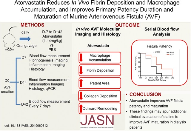
Arteriovenous fistulas placed surgically for dialysis vascular access have a high primary failure rate resulting from excessive inward remodeling, medial fibrosis, and thrombosis. No clinically established pharmacologic or perisurgical therapies currently address this unmet need. Statins' induction of multiple anti-inflammatory and antithrombotic effects suggests that these drugs might reduce arteriovenous fistula failure. Yet, the physiologic and molecular effects of statins on fistula patency and maturation remain poorly understood.
We randomized 108 C57Bl/6J mice to receive daily atorvastatin 1.14 mg/kg or PBS (control) starting 7 days before end-to-side carotid artery-jugular vein fistula creation and for up to 42 days after fistula creation. We then assessed longitudinally the effects of statin therapy on primary murine fistula patency and maturation. We concomitantly analyzed the arteriovenous fistula thrombogenic and inflammatory macrophage response to statin therapy, using the fibrin-targeted, near-infrared fluorescence molecular imaging agent FTP11-CyAm7 and dextranated, macrophage-avid nanoparticles CLIO-VT680.
molecular-structural imaging demonstrated that atorvastatin significantly reduced fibrin deposition at day 7 and macrophage accumulation at days 7 and 14, findings supported by histopathologic and gene-expression analyses. Structurally, atorvastatin promoted favorable venous limb outward remodeling, preserved arteriovenous fistula blood flow, and prolonged primary arteriovenous fistula patency through day 42 (<0.05 versus control for all measures).
These findings provide new evidence that statins improve experimental arteriovenous fistula patency and maturation, indicating that additional clinical evaluation of statin therapy in patients on dialysis undergoing arteriovenous fistula placement is warranted.
用于透析血管通路的手术置入股静脉瘘的初次通畅率很高,但由于过度内向重塑、中膜纤维化和血栓形成,导致其失败率很高。目前尚无经临床证实的药物或围手术期治疗方法可以满足这一未满足的需求。他汀类药物诱导的多种抗炎和抗血栓作用表明,这些药物可能减少动静脉瘘的失败。然而,他汀类药物对瘘管通畅性和成熟的生理和分子作用仍知之甚少。
我们将 108 只 C57Bl/6J 小鼠随机分为两组,一组每天给予阿托伐他汀 1.14mg/kg,另一组给予 PBS(对照组),从侧颈动脉-颈静脉吻合术前 7 天开始,持续 42 天。然后,我们纵向评估了他汀类药物治疗对原发性小鼠瘘管通畅性和成熟的影响。我们同时使用纤维蛋白靶向近红外荧光分子成像剂 FTP11-CyAm7 和葡聚糖化、巨噬细胞亲和性纳米颗粒 CLIO-VT680,分析了他汀类药物治疗对动静脉瘘血栓形成和炎症性巨噬细胞反应的影响。
分子结构成像显示,阿托伐他汀在第 7 天显著减少了纤维蛋白沉积,在第 7 天和第 14 天减少了巨噬细胞积累,这些发现得到了组织病理学和基因表达分析的支持。在结构上,阿托伐他汀促进了有利的静脉支向外重塑,维持了动静脉瘘的血流,并通过第 42 天延长了原发性动静脉瘘的通畅性(所有测量值均<0.05 与对照组相比)。
这些发现提供了新的证据,表明他汀类药物可改善实验性动静脉瘘的通畅性和成熟度,表明需要对接受透析治疗并进行动静脉瘘置管的患者进行他汀类药物治疗的进一步临床评估。