• 文献检索
  • 文档翻译
  • 深度研究
  • 学术资讯
  • Suppr Zotero 插件Zotero 插件
  • 邀请有礼
  • 套餐&价格
  • 历史记录
应用&插件
Suppr Zotero 插件Zotero 插件浏览器插件Mac 客户端Windows 客户端微信小程序
定价
高级版会员购买积分包购买API积分包
服务
文献检索文档翻译深度研究API 文档MCP 服务
关于我们
关于 Suppr公司介绍联系我们用户协议隐私条款
关注我们

Suppr 超能文献

核心技术专利:CN118964589B侵权必究
粤ICP备2023148730 号-1Suppr @ 2026

文献检索

告别复杂PubMed语法,用中文像聊天一样搜索,搜遍4000万医学文献。AI智能推荐,让科研检索更轻松。

立即免费搜索

文件翻译

保留排版,准确专业,支持PDF/Word/PPT等文件格式,支持 12+语言互译。

免费翻译文档

深度研究

AI帮你快速写综述,25分钟生成高质量综述,智能提取关键信息,辅助科研写作。

立即免费体验

3dMDface 和 Vectra H1 3D 面部成像系统的变异性来源。

Sources of variation in the 3dMDface and Vectra H1 3D facial imaging systems.

机构信息

Department of Anthropology, The Pennsylvania State University, University Park, PA, United States.

Department of Electrical Engineering, ESAT/PSI, KU Leuven, Leuven, Belgium.

出版信息

Sci Rep. 2020 Mar 10;10(1):4443. doi: 10.1038/s41598-020-61333-3.

DOI:10.1038/s41598-020-61333-3
PMID:32157192
原文链接:https://pmc.ncbi.nlm.nih.gov/articles/PMC7064576/
Abstract

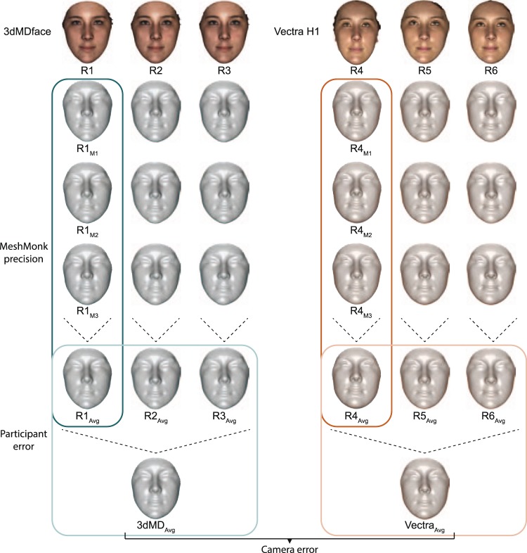
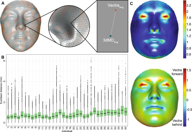
As technology advances and collaborations grow, our ability to finely quantify and explore morphological variation in 3D structures can enable important discoveries and insights into clinical, evolutionary, and genetic questions. However, it is critical to explore and understand the relative contribution of potential sources of error to the structures under study. In this study, we isolated the level of error in 3D facial images attributable to four sources, using the 3dMDface and Vectra H1 camera systems. When the two camera systems are used separately to image human participants, this analysis finds an upper bound of error potentially introduced by the use of the 3dMDface or Vectra H1 camera systems, in conjunction with the MeshMonk registration toolbox, at 0.44 mm and 0.40 mm, respectively. For studies using both camera systems, this upper bound increases to 0.85 mm, on average, and there are systematic differences in the representation of the eyelids, nostrils, and mouth by the two camera systems. Our results highlight the need for careful assessment of potential sources of error in 3D images, both in terms of magnitude and position, especially when dealing with very small measurements or performing many tests.

摘要

随着技术的进步和合作的增加,我们能够精细地定量和探索 3D 结构中的形态变化,这将为临床、进化和遗传问题的重要发现和见解提供帮助。然而,探索和理解潜在误差源对所研究结构的相对贡献至关重要。在这项研究中,我们使用 3dMDface 和 Vectra H1 相机系统,将 3D 面部图像的误差水平归因于四个来源。当分别使用两个相机系统对人类参与者进行成像时,这项分析发现,使用 3dMDface 或 Vectra H1 相机系统以及 MeshMonk 注册工具箱可能引入的误差上限分别为 0.44 毫米和 0.40 毫米。对于同时使用两个相机系统的研究,这个上限平均增加到 0.85 毫米,并且两个相机系统在眼皮、鼻孔和嘴巴的表现上存在系统差异。我们的研究结果强调了需要仔细评估 3D 图像中潜在误差源的大小和位置,尤其是在处理非常小的测量值或进行许多测试时。

https://cdn.ncbi.nlm.nih.gov/pmc/blobs/abc1/7064576/40a2adbc8ef4/41598_2020_61333_Fig4_HTML.jpg
https://cdn.ncbi.nlm.nih.gov/pmc/blobs/abc1/7064576/cfd59386cc4d/41598_2020_61333_Fig1_HTML.jpg
https://cdn.ncbi.nlm.nih.gov/pmc/blobs/abc1/7064576/b7799c16ebf4/41598_2020_61333_Fig2_HTML.jpg
https://cdn.ncbi.nlm.nih.gov/pmc/blobs/abc1/7064576/5975d5415bf1/41598_2020_61333_Fig3_HTML.jpg
https://cdn.ncbi.nlm.nih.gov/pmc/blobs/abc1/7064576/40a2adbc8ef4/41598_2020_61333_Fig4_HTML.jpg

相似文献

1
Sources of variation in the 3dMDface and Vectra H1 3D facial imaging systems.3dMDface 和 Vectra H1 3D 面部成像系统的变异性来源。
Sci Rep. 2020 Mar 10;10(1):4443. doi: 10.1038/s41598-020-61333-3.
2
Reproducibility of facial soft tissue landmarks on facial images captured on a 3D camera.3D相机拍摄的面部图像上面部软组织标志点的可重复性
Aust Orthod J. 2013 May;29(1):58-65.
3
Validation of the Vectra H1 portable three-dimensional photogrammetry system for facial imaging.用于面部成像的Vectra H1便携式三维摄影测量系统的验证
Int J Oral Maxillofac Surg. 2018 Mar;47(3):403-410. doi: 10.1016/j.ijom.2017.08.008. Epub 2017 Sep 14.
4
Digital three-dimensional photogrammetry: evaluation of anthropometric precision and accuracy using a Genex 3D camera system.数字三维摄影测量法:使用Genex 3D相机系统评估人体测量的精度和准确性。
Cleft Palate Craniofac J. 2004 Sep;41(5):507-18. doi: 10.1597/03-066.1.
5
A comparison study of different facial soft tissue analysis methods.不同面部软组织分析方法的比较研究
J Craniomaxillofac Surg. 2014 Jul;42(5):648-56. doi: 10.1016/j.jcms.2013.09.010. Epub 2013 Sep 29.
6
Craniofacial landmarks in young children: how reliable are measurements based on 3-dimensional imaging?幼儿的颅面标志点:基于三维成像的测量有多可靠?
J Craniofac Surg. 2012 Nov;23(6):1790-5. doi: 10.1097/SCS.0b013e318270fa8f.
7
Frontal soft tissue analysis using a 3 dimensional camera following two-jaw rotational orthognathic surgery in skeletal class III patients.在骨性III类患者接受双颌旋转正颌手术后,使用三维相机进行面部软组织分析。
J Craniomaxillofac Surg. 2014 Apr;42(3):220-6. doi: 10.1016/j.jcms.2013.05.004. Epub 2013 Jul 17.
8
Three-Dimensional Imaging of the Face: A Comparison Between Three Different Imaging Modalities.面部的三维成像:三种不同成像模式的比较。
Aesthet Surg J. 2018 May 15;38(6):579-585. doi: 10.1093/asj/sjx227.
9
A new method for automatic tracking of facial landmarks in 3D motion captured images (4D).一种用于在 3D 运动捕捉图像(4D)中自动跟踪面部地标点的新方法。
Int J Oral Maxillofac Surg. 2013 Jan;42(1):9-18. doi: 10.1016/j.ijom.2012.10.035. Epub 2012 Dec 4.
10
Accurate facial morphologic measurements using a 3-camera photogrammetric method.使用三相机摄影测量法进行精确的面部形态测量。
J Craniofac Surg. 2011 Jan;22(1):54-9. doi: 10.1097/SCS.0b013e3181f6c4a1.

引用本文的文献

1
Are different photogrammetry applications on smartphones sufficiently reliable?智能手机上不同的摄影测量应用程序是否足够可靠?
Korean J Orthod. 2025 Jan 25;55(1):37-47. doi: 10.4041/kjod24.134. Epub 2024 Oct 23.
2
Smartphone-generated 3D facial images: reliable for routine assessment of the oronasal region of patients with cleft or mere convenience? A validation study.智能手机生成的3D面部图像:用于唇腭裂患者口鼻区域的常规评估是可靠的还是仅仅是为了方便?一项验证研究。
BMC Oral Health. 2024 Dec 19;24(1):1517. doi: 10.1186/s12903-024-05280-9.
3
Frontiers in Three-Dimensional Surface Imaging Systems for 3D Face Acquisition in Craniofacial Research and Practice: An Updated Literature Review.

本文引用的文献

1
Comparison of Three-Dimensional Surface Imaging Systems Using Landmark Analysis.使用地标分析对三维表面成像系统进行比较
J Craniofac Surg. 2019 Sep;30(6):1869-1872. doi: 10.1097/SCS.0000000000005795.
2
MeshMonk: Open-source large-scale intensive 3D phenotyping.MeshMonk:开源大规模密集型 3D 表型分析。
Sci Rep. 2019 Apr 15;9(1):6085. doi: 10.1038/s41598-019-42533-y.
3
Six NSCL/P Loci Show Associations With Normal-Range Craniofacial Variation.六个非综合征型唇腭裂基因座与正常范围的颅面变异相关。
颅面研究与实践中用于三维面部采集的三维表面成像系统前沿:最新文献综述
Diagnostics (Basel). 2024 Feb 14;14(4):423. doi: 10.3390/diagnostics14040423.
4
Geometric learning and statistical modeling for surgical outcomes evaluation in craniosynostosis using 3D photogrammetry.基于 3D 摄影测量的颅缝早闭手术结果评估中的几何学习和统计建模。
Comput Methods Programs Biomed. 2023 Oct;240:107689. doi: 10.1016/j.cmpb.2023.107689. Epub 2023 Jun 25.
5
Validation of Vectra 3D Imaging Systems: A Review.Vectra 3D 成像系统的验证:综述
Int J Environ Res Public Health. 2022 Jul 20;19(14):8820. doi: 10.3390/ijerph19148820.
6
Decoding the Human Face: Progress and Challenges in Understanding the Genetics of Craniofacial Morphology.解码人脸:理解颅面形态遗传学的进展与挑战。
Annu Rev Genomics Hum Genet. 2022 Aug 31;23:383-412. doi: 10.1146/annurev-genom-120121-102607. Epub 2022 Apr 28.
7
Reproducibility of Novel Soft-Tissue Landmarks on Three-Dimensional Human Facial Scan Images in Caucasian and Asian.三维人脸扫描图像中新型软组织标志在白种人和亚洲人群中的可重复性。
Aesthetic Plast Surg. 2022 Apr;46(2):719-731. doi: 10.1007/s00266-021-02642-4. Epub 2021 Oct 26.
8
Large-scale open-source three-dimensional growth curves for clinical facial assessment and objective description of facial dysmorphism.大规模开源三维生长曲线用于临床面部评估和面部畸形的客观描述。
Sci Rep. 2021 Jun 9;11(1):12175. doi: 10.1038/s41598-021-91465-z.
9
3D facial phenotyping by biometric sibling matching used in contemporary genomic methodologies.通过生物特征同胞匹配进行 3D 面部表型分析,用于当代基因组方法学。
PLoS Genet. 2021 May 13;17(5):e1009528. doi: 10.1371/journal.pgen.1009528. eCollection 2021 May.
10
The Intersection of the Genetic Architectures of Orofacial Clefts and Normal Facial Variation.口腔颌面裂隙与正常面部变异的遗传结构交叉点
Front Genet. 2021 Feb 22;12:626403. doi: 10.3389/fgene.2021.626403. eCollection 2021.
Front Genet. 2018 Oct 25;9:502. doi: 10.3389/fgene.2018.00502. eCollection 2018.
4
SNPs Associated With Testosterone Levels Influence Human Facial Morphology.与睾酮水平相关的单核苷酸多态性影响人类面部形态。
Front Genet. 2018 Oct 23;9:497. doi: 10.3389/fgene.2018.00497. eCollection 2018.
5
Genome-wide mapping of global-to-local genetic effects on human facial shape.人类面部形状的全基因组范围的整体到局部遗传效应的图谱绘制。
Nat Genet. 2018 Mar;50(3):414-423. doi: 10.1038/s41588-018-0057-4. Epub 2018 Feb 19.
6
The Influence of trisomy 21 on facial form and variability.21三体对面部形态及变异性的影响。
Am J Med Genet A. 2017 Nov;173(11):2861-2872. doi: 10.1002/ajmg.a.38464. Epub 2017 Sep 21.
7
Validation of the Vectra H1 portable three-dimensional photogrammetry system for facial imaging.用于面部成像的Vectra H1便携式三维摄影测量系统的验证
Int J Oral Maxillofac Surg. 2018 Mar;47(3):403-410. doi: 10.1016/j.ijom.2017.08.008. Epub 2017 Sep 14.
8
Validation of a new three-dimensional imaging system using comparative craniofacial anthropometry.使用比较颅面人体测量法对一种新型三维成像系统进行验证。
Maxillofac Plast Reconstr Surg. 2017 Aug 25;39(1):23. doi: 10.1186/s40902-017-0123-3. eCollection 2017 Dec.
9
Evaluation of the 3dMDface system as a tool for soft tissue analysis.评估 3dMDface 系统作为软组织分析工具。
Orthod Craniofac Res. 2017 Jun;20 Suppl 1(Suppl 1):119-124. doi: 10.1111/ocr.12178.
10
Association Between Prenatal Alcohol Exposure and Craniofacial Shape of Children at 12 Months of Age.产前酒精暴露与 12 月龄儿童颅面形态的关系。
JAMA Pediatr. 2017 Aug 1;171(8):771-780. doi: 10.1001/jamapediatrics.2017.0778.