• 文献检索
  • 文档翻译
  • 深度研究
  • 学术资讯
  • Suppr Zotero 插件Zotero 插件
  • 邀请有礼
  • 套餐&价格
  • 历史记录
应用&插件
Suppr Zotero 插件Zotero 插件浏览器插件Mac 客户端Windows 客户端微信小程序
定价
高级版会员购买积分包购买API积分包
服务
文献检索文档翻译深度研究API 文档MCP 服务
关于我们
关于 Suppr公司介绍联系我们用户协议隐私条款
关注我们

Suppr 超能文献

核心技术专利:CN118964589B侵权必究
粤ICP备2023148730 号-1Suppr @ 2026

文献检索

告别复杂PubMed语法,用中文像聊天一样搜索,搜遍4000万医学文献。AI智能推荐,让科研检索更轻松。

立即免费搜索

文件翻译

保留排版,准确专业,支持PDF/Word/PPT等文件格式,支持 12+语言互译。

免费翻译文档

深度研究

AI帮你快速写综述,25分钟生成高质量综述,智能提取关键信息,辅助科研写作。

立即免费体验

SEM 下胆脂瘤侵犯人砧骨的三维图像分析归因于破骨细胞引起的骨质侵蚀,vpSEM 能谱分析揭示新骨形成。

SEM BSE 3D Image Analysis of Human Incus Bone Affected by Cholesteatoma Ascribes to Osteoclasts the Bone Erosion and VpSEM dEDX Analysis Reveals New Bone Formation.

机构信息

Department SAIMLAL Section of Human Anatomy, Laboratory of Electron Microscopy "Pietro M. Motta", Sapienza University of Rome, Via Alfonso Borelli 50, 00161 Rome, Italy.

Department NESMOS, Neurosurgery Unit, Sapienza University of Rome, Via di Grottarossa 1039, 00189 Rome, Italy.

出版信息

Scanning. 2020 Feb 15;2020:9371516. doi: 10.1155/2020/9371516. eCollection 2020.

DOI:10.1155/2020/9371516
PMID:32158510
原文链接:https://pmc.ncbi.nlm.nih.gov/articles/PMC7048945/
Abstract

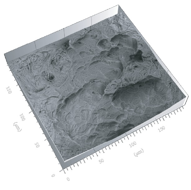
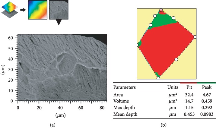
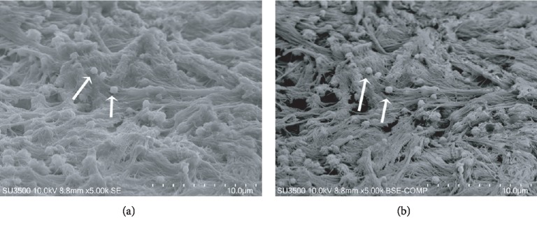
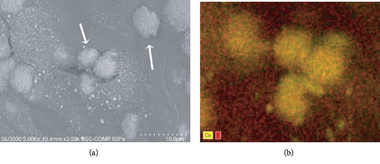
Bone erosion is considered a typical characteristic of advanced or complicated cholesteatoma (CHO), although it is still a matter of debate if bone erosion is due to osteoclast action, being the specific literature controversial. The purpose of this study was to apply a novel scanning characterization approach, the BSE 3D image analysis, to study the pathological erosion on the surface of human incus bone involved by CHO, in order to definitely assess the eventual osteoclastic resorptive action. To do this, a comparison of BSE 3D image of resorption lacunae (resorption pits) from osteoporotic human femur neck (indubitably of osteoclastic origin) with that of the incus was performed. Surface parameters (area, mean depth, and volume) were calculated by the software Hitachi MountainsMap© from BSE 3D-reconstructed images; results were then statistically analyzed by SPSS statistical software. Our findings showed that no significant differences exist between the two groups. This quantitative approach implements the morphological characterization, allowing us to state that surface erosion of the incus is due to osteoclast action. Moreover, our observation and processing image workflow are the first in the literature showing the presence not only of bone erosion but also of matrix vesicles releasing their content on collagen bundles and self-immuring osteocytes, all markers of new bone formation on incus bone surface. On the basis of recent literature, it has been hypothesized that inflammatory environment induced by CHO may trigger the osteoclast activity, eliciting bone erosion. The observed new bone formation probably takes place at a slower rate in respect to the normal bone turnover, and the process is uncoupled (as recently demonstrated for several inflammatory diseases that promote bone loss) thus resulting in an overall bone loss. Novel scanning characterization approaches used in this study allowed for the first time the 3D imaging of incus bone erosion and its quantitative measurement, opening a new era of quantitative SEM morphology.

摘要

骨质侵蚀被认为是高级或复杂胆脂瘤(CHO)的典型特征,尽管骨质侵蚀是否是由于破骨细胞的作用导致,仍然存在争议,具体文献存在争议。本研究旨在应用一种新的扫描特征化方法,即 BSE 3D 图像分析,来研究 CHO 累及的人砧骨表面的病理性侵蚀,以明确评估潜在的破骨细胞吸收作用。为此,对骨质疏松性人股骨颈的吸收陷窝(吸收坑)的 BSE 3D 图像(无疑是破骨细胞起源)与砧骨的 BSE 3D 图像进行了比较。通过 Hitachi MountainsMap©软件从 BSE 3D 重建图像中计算表面参数(面积、平均深度和体积);然后使用 SPSS 统计软件对结果进行统计学分析。我们的发现表明,两组之间没有显著差异。这种定量方法实现了形态特征化,使我们能够断言砧骨的表面侵蚀是由破骨细胞作用引起的。此外,我们的观察和图像处理工作流程是文献中首次显示不仅存在骨质侵蚀,还存在基质小泡释放其内容物到胶原束上并自我封闭成骨细胞,所有这些都是砧骨表面新骨形成的标志物。根据最近的文献,有人假设 CHO 引起的炎症环境可能会触发破骨细胞活性,引发骨质侵蚀。观察到的新骨形成可能以比正常骨转换更慢的速度发生,并且该过程是解偶联的(如最近在几种促骨丢失的炎症性疾病中所证明的),从而导致整体骨丢失。本研究中使用的新型扫描特征化方法首次允许对砧骨骨质侵蚀进行 3D 成像及其定量测量,开创了定量 SEM 形态学的新时代。

https://cdn.ncbi.nlm.nih.gov/pmc/blobs/0901/7048945/c0177bba19da/SCANNING2020-9371516.010.jpg
https://cdn.ncbi.nlm.nih.gov/pmc/blobs/0901/7048945/f34c2d0c08b9/SCANNING2020-9371516.001.jpg
https://cdn.ncbi.nlm.nih.gov/pmc/blobs/0901/7048945/dd130ad09d39/SCANNING2020-9371516.002.jpg
https://cdn.ncbi.nlm.nih.gov/pmc/blobs/0901/7048945/6fe6bff76f62/SCANNING2020-9371516.003.jpg
https://cdn.ncbi.nlm.nih.gov/pmc/blobs/0901/7048945/93d65f2e5b4e/SCANNING2020-9371516.004.jpg
https://cdn.ncbi.nlm.nih.gov/pmc/blobs/0901/7048945/95c6f6e15afd/SCANNING2020-9371516.005.jpg
https://cdn.ncbi.nlm.nih.gov/pmc/blobs/0901/7048945/4c1362aac634/SCANNING2020-9371516.006.jpg
https://cdn.ncbi.nlm.nih.gov/pmc/blobs/0901/7048945/1c84036b513a/SCANNING2020-9371516.007.jpg
https://cdn.ncbi.nlm.nih.gov/pmc/blobs/0901/7048945/ddd38952c884/SCANNING2020-9371516.008.jpg
https://cdn.ncbi.nlm.nih.gov/pmc/blobs/0901/7048945/30a959b77806/SCANNING2020-9371516.009.jpg
https://cdn.ncbi.nlm.nih.gov/pmc/blobs/0901/7048945/c0177bba19da/SCANNING2020-9371516.010.jpg

相似文献

1
SEM BSE 3D Image Analysis of Human Incus Bone Affected by Cholesteatoma Ascribes to Osteoclasts the Bone Erosion and VpSEM dEDX Analysis Reveals New Bone Formation.SEM 下胆脂瘤侵犯人砧骨的三维图像分析归因于破骨细胞引起的骨质侵蚀,vpSEM 能谱分析揭示新骨形成。
Scanning. 2020 Feb 15;2020:9371516. doi: 10.1155/2020/9371516. eCollection 2020.
2
Osteoclasts in chronic otitis media, cholesteatoma, and otosclerosis.慢性中耳炎、胆脂瘤和耳硬化症中的破骨细胞。
Ann Otol Rhinol Laryngol. 1988 Nov-Dec;97(6 Pt 1):661-6. doi: 10.1177/000348948809700615.
3
Measuring mineralised tissue formation and resorption in a human 3D osteoblast-osteoclast co-culture model.测量人源成骨细胞-破骨细胞共培养 3D 模型中的矿化组织形成和吸收。
Eur Cell Mater. 2020 Nov 5;40:189-202. doi: 10.22203/eCM.v040a12.
4
Cellular and subcellular events of bone resorption in human and experimental cholesteatoma: the role of osteoclasts.人类及实验性胆脂瘤中骨吸收的细胞及亚细胞事件:破骨细胞的作用
Laryngoscope. 1984 Jan;94(1):76-95. doi: 10.1002/lary.5540940117.
5
Erosion of the incus in pediatric posterior tympanic membrane retraction pockets without cholesteatoma.小儿无胆脂瘤型后鼓膜内陷袋中砧骨侵蚀
Int J Pediatr Otorhinolaryngol. 2008 Sep;72(9):1419-23. doi: 10.1016/j.ijporl.2008.06.004. Epub 2008 Jul 16.
6
Steering the osteoclast through the demineralization-collagenolysis balance.引导破骨细胞通过脱矿质-胶原溶解平衡。
Bone. 2013 Sep;56(1):191-8. doi: 10.1016/j.bone.2013.06.007. Epub 2013 Jun 15.
7
Bone Resorption Activity in Mature Osteoclasts.成熟破骨细胞中的骨吸收活性。
Methods Mol Biol. 2018;1868:215-222. doi: 10.1007/978-1-4939-8802-0_22.
8
Higher degree of keratinization correlated with severe bone destruction in acquired Cholesteatoma.角化程度越高,获得性胆脂瘤的骨破坏越严重。
Acta Otolaryngol. 2023 Feb;143(2):147-155. doi: 10.1080/00016489.2023.2177341. Epub 2023 Mar 8.
9
Inhibition of osteoclast differentiation and collagen antibody-induced arthritis by CTHRC1.CTHRC1对破骨细胞分化及胶原抗体诱导性关节炎的抑制作用
Bone. 2017 Apr;97:153-167. doi: 10.1016/j.bone.2017.01.022. Epub 2017 Jan 21.
10
Activation of dimeric glucocorticoid receptors in osteoclast progenitors potentiates RANKL induced mature osteoclast bone resorbing activity.破骨细胞前体细胞中二聚体糖皮质激素受体的激活增强了RANKL诱导的成熟破骨细胞的骨吸收活性。
Bone. 2016 Dec;93:43-54. doi: 10.1016/j.bone.2016.08.024. Epub 2016 Sep 2.

引用本文的文献

1
Improved Cholesteatoma Removal with CADISS: A Quantitative Ultrastructural Comparison Using VP-SEM.使用CADISS改善胆脂瘤切除:基于可变压力扫描电子显微镜的定量超微结构比较
J Clin Med. 2025 Jun 12;14(12):4192. doi: 10.3390/jcm14124192.
2
PTHrP participates in the bone destruction of middle ear cholesteatoma via promoting macrophage differentiation into osteoclasts induced by RANKL.甲状旁腺激素相关蛋白通过促进破骨细胞分化诱导因子 RANKL 促进中耳胆脂瘤骨破坏。
Zhong Nan Da Xue Xue Bao Yi Xue Ban. 2024 May 28;49(5):655-666. doi: 10.11817/j.issn.1672-7347.2024.230482.
3
Biological Effects of Small Sized Graphene Oxide Nanosheets on Human Leukocytes.

本文引用的文献

1
Osteoclasts Modulate Bone Erosion in Cholesteatoma via RANKL Signaling.破骨细胞通过 RANKL 信号调节胆脂瘤中的骨质侵蚀。
J Assoc Res Otolaryngol. 2019 Oct;20(5):449-459. doi: 10.1007/s10162-019-00727-1. Epub 2019 Jun 28.
2
Ciliated cell observation by SEM on the surface of human incudo-malleolar-joint articular cartilage: are they a new chondrocyte phenotype?通过扫描电子显微镜对人砧镫关节软骨表面纤毛细胞的观察:它们是一种新的软骨细胞表型吗?
Acta Otolaryngol. 2019 May;139(5):439-443. doi: 10.1080/00016489.2019.1575520. Epub 2019 Feb 26.
3
Scanning Electron Microscopy of Bone.
小尺寸氧化石墨烯纳米片对人白细胞的生物学效应
Biomedicines. 2024 Jan 23;12(2):256. doi: 10.3390/biomedicines12020256.
4
Cholesteatoma Severely Impacts the Integrity and Bone Material Quality of the Incus.胆脂瘤严重影响砧骨的完整性和骨质质量。
Calcif Tissue Int. 2023 Dec;113(6):609-617. doi: 10.1007/s00223-023-01144-6. Epub 2023 Oct 24.
5
Adult IDH Wild-Type Glioblastoma Ultrastructural Investigation Suggests a Possible Correlation between Morphological Biomarkers and Ki-67 Index.成人异柠檬酸脱氢酶野生型胶质母细胞瘤的超微结构研究表明形态学生物标志物与Ki-67指数之间可能存在相关性。
Biomedicines. 2023 Jul 12;11(7):1968. doi: 10.3390/biomedicines11071968.
6
The Application of a Fluoride-and-Vitamin D Solution to Deciduous Teeth Promotes Formation of Persistent Mineral Crystals: A Morphological Ex-Vivo Study.氟化物与维生素D溶液应用于乳牙促进持久性矿物晶体形成:一项形态学体外研究
Materials (Basel). 2023 May 29;16(11):4049. doi: 10.3390/ma16114049.
7
Effect of Ciprofloxacin-Loaded Niosomes on and Biofilm Formation.载环丙沙星脂质体对[具体内容缺失]和生物膜形成的影响。
Pharmaceutics. 2022 Nov 30;14(12):2662. doi: 10.3390/pharmaceutics14122662.
8
Anti-Virulence Potential of a Chionodracine-Derived Peptide against Multidrug-Resistant Clinical Isolates from Cystic Fibrosis Patients.抗真菌肽对囊性纤维化患者多药耐药临床分离株的抗毒力作用
Int J Mol Sci. 2022 Nov 4;23(21):13494. doi: 10.3390/ijms232113494.
9
A Different Exosome Secretion Pattern Characterizes Patient-Derived Colorectal Cancer Multicellular Spheroids and Their Mouse Xenografts.不同的外泌体分泌模式是患者来源的结直肠癌多细胞球体及其小鼠异种移植瘤的特征。
Biology (Basel). 2022 Sep 29;11(10):1427. doi: 10.3390/biology11101427.
10
Review of potential medical treatments for middle ear cholesteatoma.中耳胆脂瘤潜在医学治疗方法的综述。
Cell Commun Signal. 2022 Sep 19;20(1):148. doi: 10.1186/s12964-022-00953-w.
骨的扫描电子显微镜检查
Methods Mol Biol. 2019;1914:571-616. doi: 10.1007/978-1-4939-8997-3_31.
4
Bone Loss in Rheumatoid Arthritis: Basic Mechanisms and Clinical Implications.类风湿关节炎的骨丢失:基础机制与临床意义。
Calcif Tissue Int. 2018 May;102(5):533-546. doi: 10.1007/s00223-017-0373-1. Epub 2017 Dec 4.
5
Inflammatory osteolysis: a conspiracy against bone.炎性骨溶解:一场针对骨骼的“阴谋”。
J Clin Invest. 2017 Jun 1;127(6):2030-2039. doi: 10.1172/JCI93356.
6
The role of bone resorption in the etiopathogenesis of acquired middle ear cholesteatoma.骨吸收在后天性中耳胆脂瘤发病机制中的作用。
Eur Arch Otorhinolaryngol. 2017 May;274(5):2071-2078. doi: 10.1007/s00405-016-4422-6. Epub 2016 Dec 16.
7
Presence of osteoclasts in middle ear cholesteatoma: a study of undecalcified bone sections.中耳胆脂瘤中破骨细胞的存在:对未脱钙骨切片的研究
Acta Otolaryngol. 2017 Feb;137(2):127-130. doi: 10.1080/00016489.2016.1222549. Epub 2016 Aug 30.
8
Intercellular Communication between Keratinocytes and Fibroblasts Induces Local Osteoclast Differentiation: a Mechanism Underlying Cholesteatoma-Induced Bone Destruction.角质形成细胞与成纤维细胞之间的细胞间通讯诱导局部破骨细胞分化:胆脂瘤诱导骨破坏的潜在机制。
Mol Cell Biol. 2016 May 16;36(11):1610-20. doi: 10.1128/MCB.01028-15. Print 2016 Jun 1.
9
Bacterial Infections and Osteoclastogenesis Regulators in Men and Women with Cholesteatoma.胆脂瘤患者中男性和女性的细菌感染与破骨细胞生成调节剂
Arch Immunol Ther Exp (Warsz). 2016 Jun;64(3):241-7. doi: 10.1007/s00005-015-0373-7. Epub 2015 Nov 19.
10
Biological Niches within Human Calcified Aortic Valves: Towards Understanding of the Pathological Biomineralization Process.人类钙化主动脉瓣内的生物龛:迈向对病理性生物矿化过程的理解
Biomed Res Int. 2015;2015:542687. doi: 10.1155/2015/542687. Epub 2015 Oct 5.