MINTOS research group - Medical Imaging and Navigation in Trauma and Orthopedic Surgery, Department for Trauma and Orthopedic Surgery, BG Trauma Center Ludwigshafen at Heidelberg University Hospital, Ludwig-Guttmann-Str. 13, 67071, Ludwigshafen, Germany.
AGiTEC - Working Group for intraoperative imaging and integration of technologies of the DGOU, Berlin, Germany.
Sci Rep. 2020 Mar 11;10(1):4530. doi: 10.1038/s41598-020-61267-w.
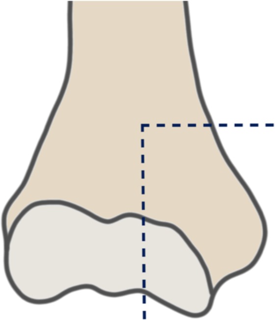
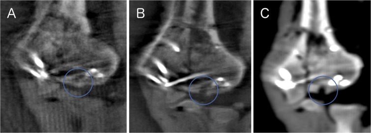
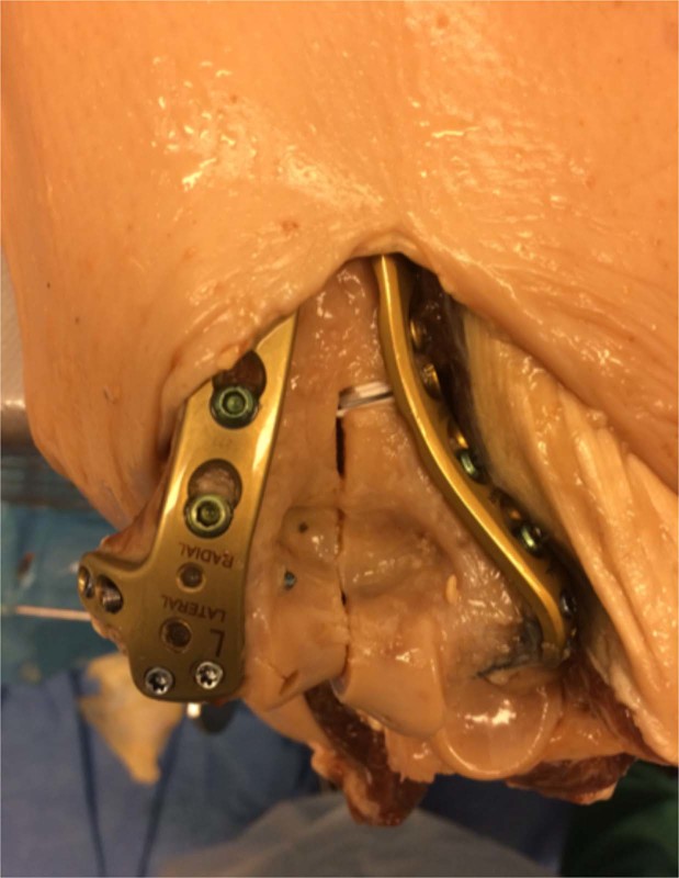
Anatomic reconstruction of articular fractures is one of the critical factors in later achieving good functional outcome. Intraoperative 3D imaging has been shown to offer better evaluation and therefore can significantly improve the results. The purpose of this study was to assess the difference between intraoperative three-dimensional fluoroscopy (3D) and intraoperative computed tomography (iCT) imaging regarding fracture reduction, implant placement and articular impressions in a distal humeral fracture model. AO type 13-B2 fracture pattern were created in upper extremity cadaver specimens. Articular step-offs, intra-articular screw placement and intraarticular impressions of different degrees of severity were created. All specimens had imaging performed. For each articular pattern 3D fluoroscopy in standard (3Ds) and high quality (3Dh) were performed (Arcadis Orbic, Siemens, Germany) as well as an intraoperative CT scan (iCT, Airo, Brainlab, Germany). Three observers evaluated all imaging studies regarding subjective and objective parameters. iCT is more precise than 3D fluoroscopic imaging for detection of articular impressions. Articular step-offs and intraarticular screw placement are similar for iCT and 3D. Subjective imaging quality is the highest for iCT and lowest for 3Ds. Intraoperative CT may be particularly useful in assessing articular impressions and providing a good subjective image quality for the surgeon.
关节骨折的解剖重建是获得良好功能结果的关键因素之一。术中三维成像已被证明可提供更好的评估,因此可以显著改善结果。本研究旨在评估在肱骨远端骨折模型中,术中三维透视(3D)与术中计算机断层扫描(iCT)成像在骨折复位、植入物放置和关节压痕方面的差异。在上肢尸体标本上创建了 AO 13-B2 型骨折模式。创建了关节台阶、关节内螺钉放置和不同严重程度的关节内压痕。所有标本均进行了影像学检查。对于每个关节形态,均进行了标准(3Ds)和高质量(3Dh)的三维透视(Arcadis Orbic,Siemens,德国)以及术中 CT 扫描(iCT,Airo,Brainlab,德国)。三位观察者根据主观和客观参数评估所有影像学研究。iCT 比 3D 透视成像更能准确检测关节压痕。关节台阶和关节内螺钉放置在 iCT 和 3D 之间相似。iCT 的主观成像质量最高,3Ds 的最低。术中 CT 可能特别有助于评估关节压痕,并为外科医生提供良好的主观图像质量。