Université de Paris, BIOSCAR Inserm U1132 and Department of Rheumatology and Reference Center for Constitutional Bone Diseases, AP-HP Hospital Lariboisière, F-75010, Paris, France.
Université de Paris, Department of Bone and Joint Imaging, AP-HP Hospital Lariboisière, F-75010, Paris, France.
Sci Rep. 2020 Mar 13;10(1):4699. doi: 10.1038/s41598-020-61704-w.
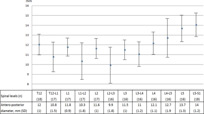
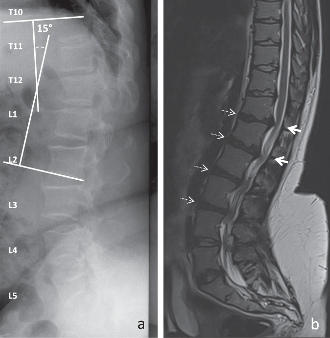
In achondroplasia, lumbar spinal stenosis arises from congenital dysplasia and acquired degenerative changes. We here aimed to describe the changes of the lumbar spinal canal and intervertebral disc in adults. We included 18 adults (age ≥ 18 years) with achondroplasia and lumbar spinal stenosis. Radiographs were used to analyze spinal-pelvic angles. Antero-posterior diameter of the spinal canal and the grade of disc degeneration were measured by MRI. Antero-posterior diameters of the spinal canal differed by spinal level (P < 0.05), with lower values observed at T12-L1, L1-2 and L2-3. Degrees of disc degeneration differed by intervertebral level, with higher degrees observed at L1-2, L2-3 and L3-4. A significant correlation was found between disc degeneration and thoraco-lumbar kyphosis at L2-3, between antero-posterior diameter of the spinal canal and lumbar lordosis at T12-L1 and L2-3, and between antero-posterior diameter of the spinal canal and thoraco-lumbar kyphosis at L1-2. Unlike the general population, spinal stenosis and disc degeneration involve the upper part of the lumbar spine in adults with achondroplasia, associated with thoraco-lumbar kyphosis and loss of lumbar lordosis.
在软骨发育不全中,腰椎管狭窄症是由先天性发育不良和获得性退行性改变引起的。我们旨在描述成年人腰椎管和椎间盘的变化。我们纳入了 18 名患有软骨发育不全和腰椎管狭窄症的成年人(年龄≥18 岁)。通过 X 线片分析脊柱骨盆角。通过 MRI 测量椎管前后径和椎间盘退变程度。椎管前后径随脊柱水平而变化(P<0.05),T12-L1、L1-2 和 L2-3 水平较低。椎间盘退变程度随椎间水平而变化,L1-2、L2-3 和 L3-4 水平较高。在 L2-3 水平,椎间盘退变与胸腰椎后凸之间存在显著相关性;在 T12-L1 和 L2-3 水平,椎管前后径与腰椎前凸之间存在显著相关性;在 L1-2 水平,椎管前后径与胸腰椎后凸之间存在显著相关性。与一般人群不同,患有软骨发育不全的成年人的腰椎管狭窄症和椎间盘退变累及腰椎上段,与胸腰椎后凸和腰椎前凸丢失有关。