Department of Respiratory Medicine and Center for Translational Immunology, University Medical Center Utrecht, The Netherlands.
Department of Surgery, University Medical Center Utrecht, The Netherlands.
J Leukoc Biol. 2020 May;107(5):809-818. doi: 10.1002/JLB.5HR0120-459R. Epub 2020 Mar 14.
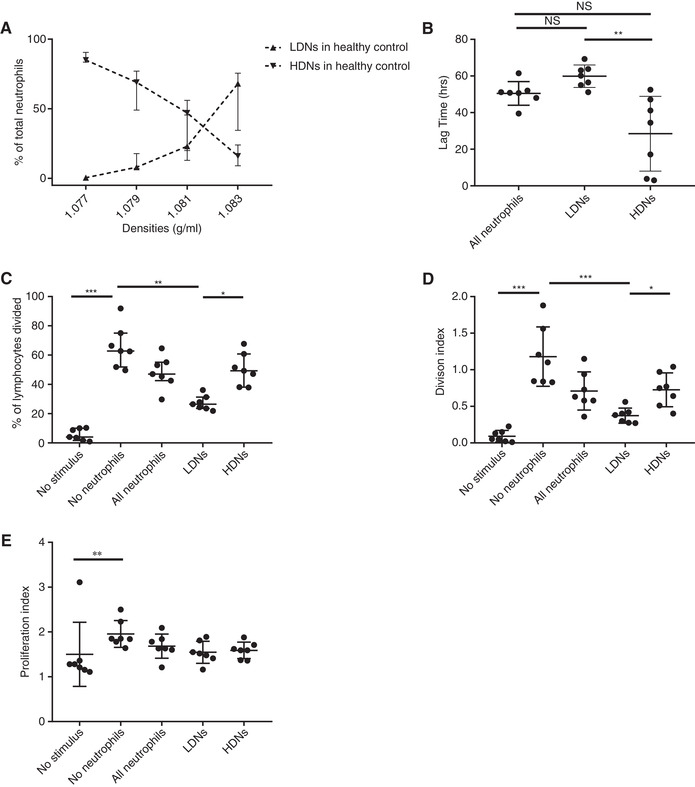
Here we elaborate on the origin of low(er)-density neutrophils (LDNs) to better understand the variation found in literature. Supplemented with original data, we test the hypothesis that buoyant density of neutrophils is characterized by a spectrum that as a whole shifts to a lower density after activation. Both the 20% highest density (HDNs) and 20% lowest density (LDNs) neutrophils from healthy donors were isolated by Percoll of different densities. Using this method we found that LDNs were significantly better in T-cell suppression and bacterial containment than their 20% highest density counterparts. We found no statistically relevant differences in neutrophil survival or bacterial phagocytosis. Stimulation of healthy donor neutrophils with N-formyl-methionyl-leucyl-phenylalanine induced LDNs co-segregating with peripheral blood mononuclear cells after Ficoll separation. These in vitro induced LDNs showed increased activation markers compared to HDNs and were comparable to the activation markers found on the LDN fraction seen in patients with chronic inflammatory conditions such as present in cancer patients. This all fits with the hypothesis that the density of neutrophils is distributed in a spectrum partially coupled to maturation. Additionally a shift in this spectrum can be induced by in vitro stimulation or by activation in disease.
在这里,我们详细阐述了低(或)密度中性粒细胞(LDN)的起源,以便更好地理解文献中发现的变化。我们补充了原始数据,检验了这样一个假设,即中性粒细胞的浮力密度具有一个整体向较低密度移动的谱,这种移动发生在激活之后。通过不同密度的 Percoll,从健康供体中分离出 20%最高密度(HDNs)和 20%最低密度(LDNs)中性粒细胞。使用这种方法,我们发现 LDNs 在抑制 T 细胞和控制细菌方面明显优于其 20%最高密度的对应物。我们没有发现中性粒细胞存活或细菌吞噬方面存在统计学上显著的差异。用 N-甲酰基-甲硫氨酸-亮氨酸-苯丙氨酸刺激健康供体中性粒细胞,在 Ficoll 分离后,LDN 与外周血单核细胞共同分离。与 HDNs 相比,这些体外诱导的 LDNs 显示出更高的激活标志物,与在患有慢性炎症性疾病(如癌症患者中)的患者中看到的 LDN 部分相关的激活标志物相当。这一切都符合这样一个假设,即中性粒细胞的密度分布在一个与成熟部分相关的谱中。此外,这种谱的移动可以通过体外刺激或疾病激活来诱导。