Center for Reproductive Medicine, Research Institute Reproduction and Development, Amsterdam University Medical Center, University of Amsterdam, Amsterdam, The Netherlands.
Department of Pathology, Erasmus MC University Medical Center, Rotterdam, and Princess Maxima Center for Pediatric Oncology, Utrecht, The Netherlands.
PLoS One. 2020 Mar 16;15(3):e0230253. doi: 10.1371/journal.pone.0230253. eCollection 2020.
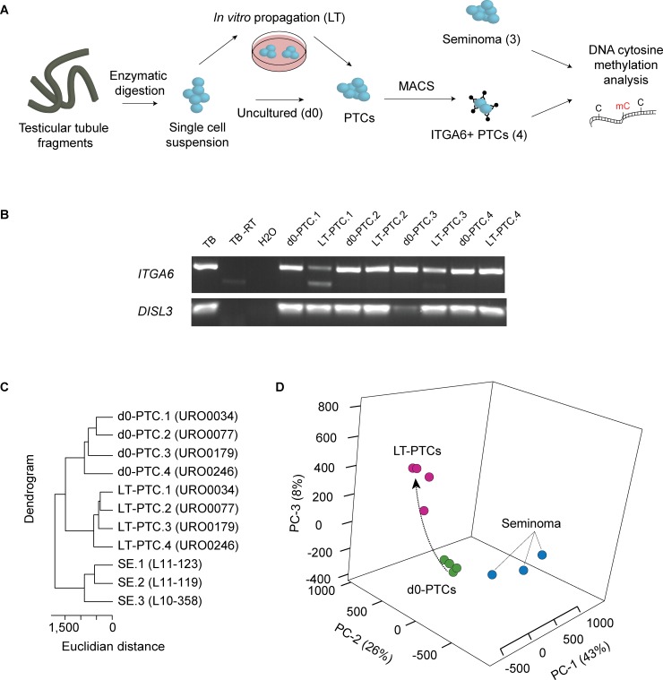
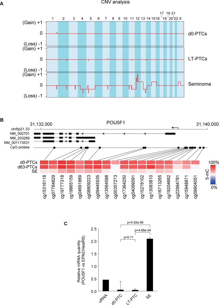
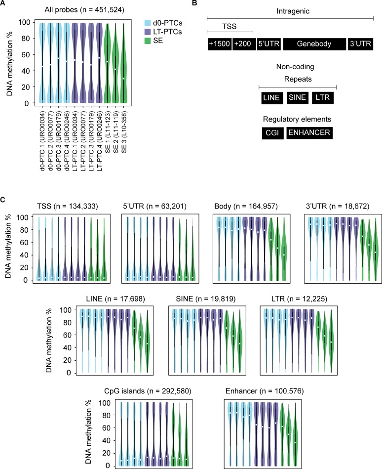
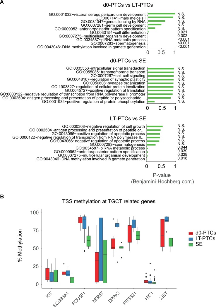
Autologous transplantation of spermatogonial stem cells is a promising new avenue to restore fertility in infertile recipients. Expansion of the initial spermatogonial stem cell pool through cell culturing is a necessary step to obtain enough cells for effective repopulation of the testis after transplantation. Since in vitro propagation can lead to (epi-)genetic mutations and possibly malignant transformation of the starting cell population, we set out to investigate genome-wide DNA methylation status in uncultured and cultured primary testicular ITGA6+ sorted cells and compare them with germ cell tumor samples of the seminoma subtype. Seminomas displayed a severely global hypomethylated profile, including loss of genomic imprinting, which we did not detect in cultured primary testicular ITGA6+ cells. Differential methylation analysis revealed altered regulation of gamete formation and meiotic processes in cultured primary testicular ITGA6+ cells but not in seminomas. The pivotal POU5F1 marker was hypomethylated in seminomas but not in uncultured or cultured primary testicular ITGA6+ cells, which is reflected in the POU5F1 mRNA expression levels. Lastly, seminomas displayed a number of characteristic copy number variations that were not detectable in primary testicular ITGA6+ cells, either before or after culture. Together, the data show a distinct DNA methylation patterns in cultured primary testicular ITGA6+ cells that does not resemble the pattern found in seminomas, but also highlight the need for more sensitive methods to fully exclude the presence of malignant cells after culture and to further study the epigenetic events that take place during in vitro culture.
自体移植精原干细胞是恢复不育受者生育能力的一种有前途的新途径。通过细胞培养来扩大初始精原干细胞池是获得足够细胞以有效重 populate 移植后睾丸的必要步骤。由于体外繁殖可能导致( epi )遗传突变,并可能导致起始细胞群体的恶性转化,因此我们着手研究未培养和培养的原发性睾丸 ITGA6+分选细胞的全基因组 DNA 甲基化状态,并将其与精原细胞瘤样本进行比较。精原细胞瘤表现出严重的全局低甲基化谱,包括基因组印记的丧失,而我们在未培养和培养的原发性睾丸 ITGA6+细胞中未检测到这种情况。差异甲基化分析显示,培养的原发性睾丸 ITGA6+细胞中配子形成和减数分裂过程的调节发生改变,但在精原细胞瘤中未发现这种情况。关键的 POU5F1 标记在精原细胞瘤中被低甲基化,但在未培养或培养的原发性睾丸 ITGA6+细胞中未被检测到,这反映在 POU5F1 mRNA 表达水平上。最后,精原细胞瘤显示出许多特征性的拷贝数变异,这些变异在原发性睾丸 ITGA6+细胞中无论是在培养之前还是之后都无法检测到。总之,这些数据显示培养的原发性睾丸 ITGA6+细胞中的 DNA 甲基化模式与精原细胞瘤中发现的模式明显不同,但也强调需要更敏感的方法来完全排除培养后恶性细胞的存在,并进一步研究体外培养过程中发生的表观遗传事件。