Pathological Anatomy Center of University Hospital of Brasilia, Via L2 Norte, SGAN 604/605, Brasília DF, 70840-050, Brazil.
Pathology Department of Brasília University, Brasília, Brazil.
BMC Cancer. 2020 Mar 17;20(1):225. doi: 10.1186/s12885-020-6670-5.
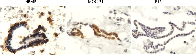
Considering the potential of p16 as a marker for diagnosis, prognosis and therapeutic response, the aim of this study was to assess its presence, via immunocytochemistry, in metastatic carcinoma of different primary sites and histological types obtained from effusions and peritoneal washings. A total of 118 samples including 85 of metastatic carcinoma and 33 samples of benign effusion/peritoneal washing were prepared by the plasma/thromboplastin method. Immunocytochemistry reactions were performed on cell block sections using antibodies against p16, claudin-4, MOC-31, calretinin, HBME and CD68.
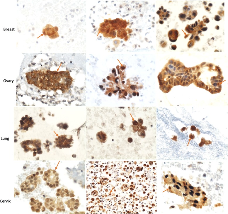
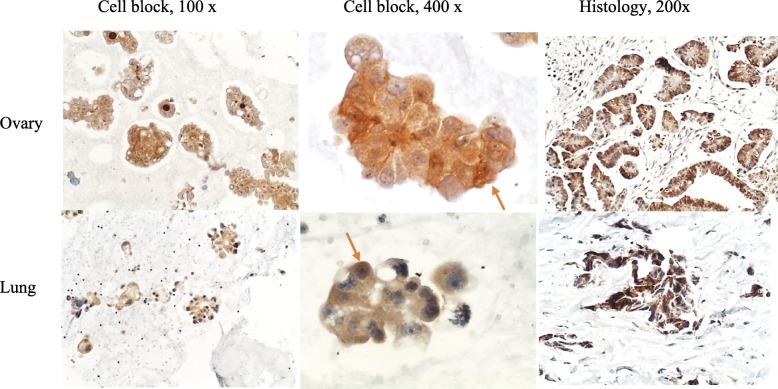
P16 overexpression was observed in 88.23% of all carcinoma samples. All cervix adenocarcinoma samples showed p16 overexpression. Overexpression in adenocarcinomas of ovary, lung and breast was observed in 93.75, 93.10 and 75% of the samples, respectively. Overexpression was observed in all different histological types analyzed: small cell carcinoma (lung), squamous cell carcinoma (cervical) and urothelial carcinoma (bladder). The specificity of p16 for carcinoma detection was of 96.96%.
Overexpression of p16 was observed in most metastatic carcinoma, from different primary sites and histological types, obtained from effusions and peritoneal washings. Due to its high frequency of overexpression in metastatic carcinoma, p16 may play a possible role in tumor progression and it may be considered as a complementary diagnostic marker depending on histological type and primary site of carcinoma.
鉴于 p16 作为诊断、预后和治疗反应标志物的潜力,本研究旨在通过免疫细胞化学评估其在来自胸腔积液和腹腔冲洗液的不同原发部位和组织学类型的转移性癌中的存在。共制备了 118 个样本,包括 85 个转移性癌样本和 33 个良性胸腔积液/腹腔冲洗液样本,通过血浆/凝血酶原方法制备。使用针对 p16、claudin-4、MOC-31、钙视网膜蛋白、HBME 和 CD68 的抗体对细胞块切片进行免疫细胞化学反应。
所有癌样本中 p16 过表达率为 88.23%。所有宫颈腺癌样本均显示 p16 过表达。卵巢、肺和乳腺腺癌的过表达率分别为 93.75%、93.10%和 75%。在分析的所有不同组织学类型中均观察到过表达:小细胞癌(肺)、鳞状细胞癌(宫颈)和尿路上皮癌(膀胱)。p16 对癌检测的特异性为 96.96%。
在来自胸腔积液和腹腔冲洗液的不同原发部位和组织学类型的大多数转移性癌中观察到 p16 过表达。由于其在转移性癌中的过表达频率较高,p16 可能在肿瘤进展中发挥作用,并可能根据癌的组织学类型和原发部位被视为补充诊断标志物。