• 文献检索
  • 文档翻译
  • 深度研究
  • 学术资讯
  • Suppr Zotero 插件Zotero 插件
  • 邀请有礼
  • 套餐&价格
  • 历史记录
应用&插件
Suppr Zotero 插件Zotero 插件浏览器插件Mac 客户端Windows 客户端微信小程序
定价
高级版会员购买积分包购买API积分包
服务
文献检索文档翻译深度研究API 文档MCP 服务
关于我们
关于 Suppr公司介绍联系我们用户协议隐私条款
关注我们

Suppr 超能文献

核心技术专利:CN118964589B侵权必究
粤ICP备2023148730 号-1Suppr @ 2026

文献检索

告别复杂PubMed语法,用中文像聊天一样搜索,搜遍4000万医学文献。AI智能推荐,让科研检索更轻松。

立即免费搜索

文件翻译

保留排版,准确专业,支持PDF/Word/PPT等文件格式,支持 12+语言互译。

免费翻译文档

深度研究

AI帮你快速写综述,25分钟生成高质量综述,智能提取关键信息,辅助科研写作。

立即免费体验

腹腔内的脂肪异常:一篇图文并茂的文章。

Fat misbehaving in the abdominal cavity: a pictorial essay.

作者信息

Devos Hannes, Goethals Lode, Belsack Dries, Brucker Yannick De, Allemeersch Gert-Jan, Ilsen Bart, Vandenbroucke Frederik, de Mey Johan

机构信息

UZ Brussel, Belgium.

出版信息

Pol J Radiol. 2020 Jan 21;85:e32-e38. doi: 10.5114/pjr.2020.93070. eCollection 2020.

DOI:10.5114/pjr.2020.93070
PMID:32180852
原文链接:https://pmc.ncbi.nlm.nih.gov/articles/PMC7064015/
Abstract

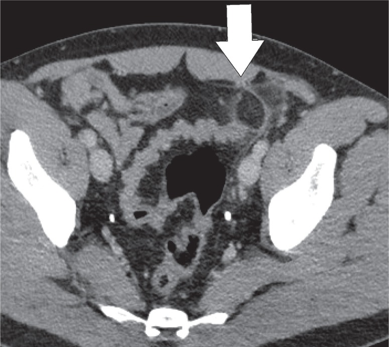
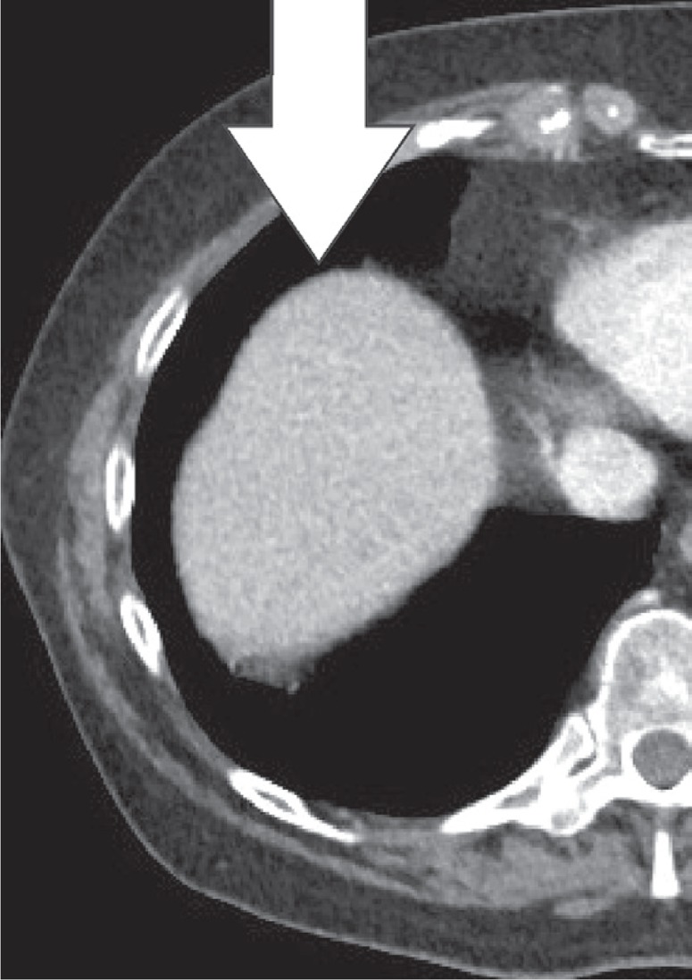
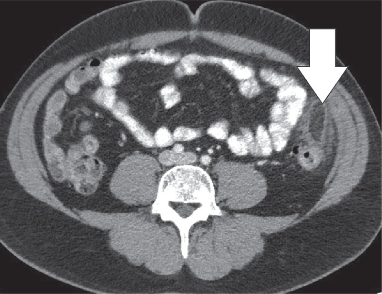
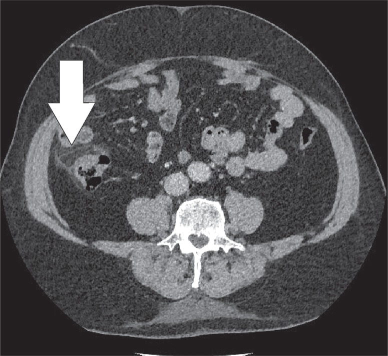
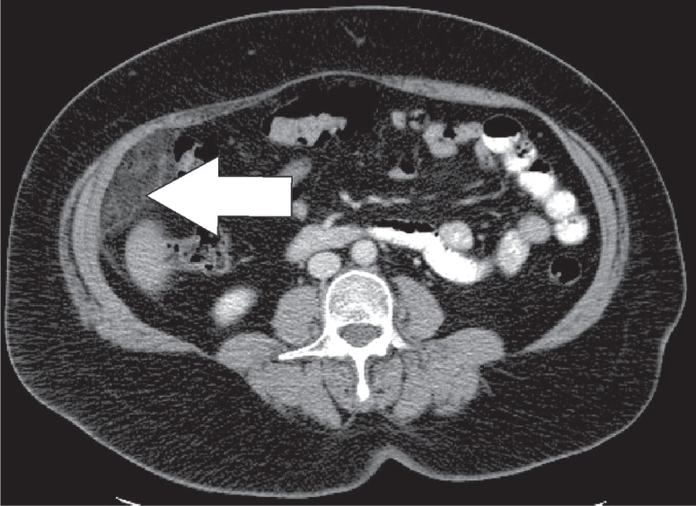
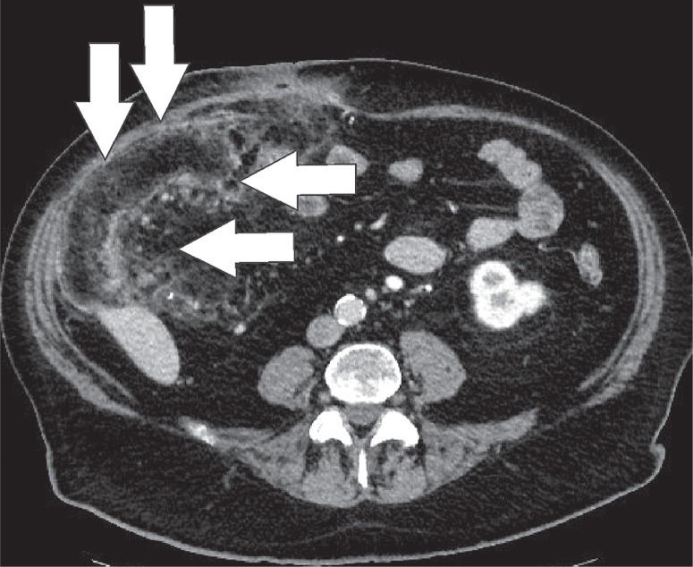
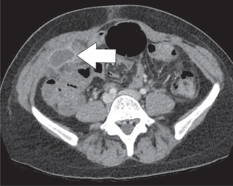
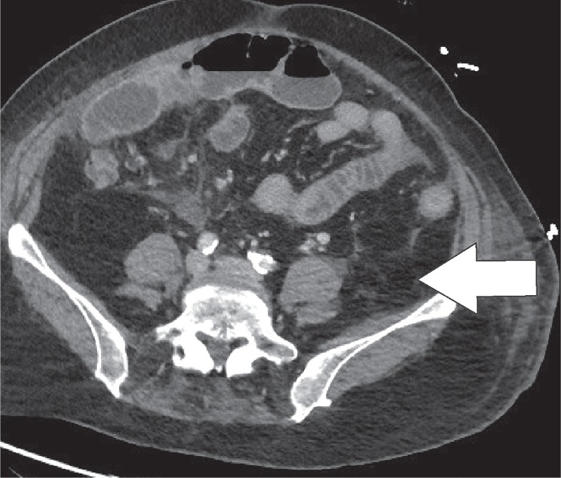
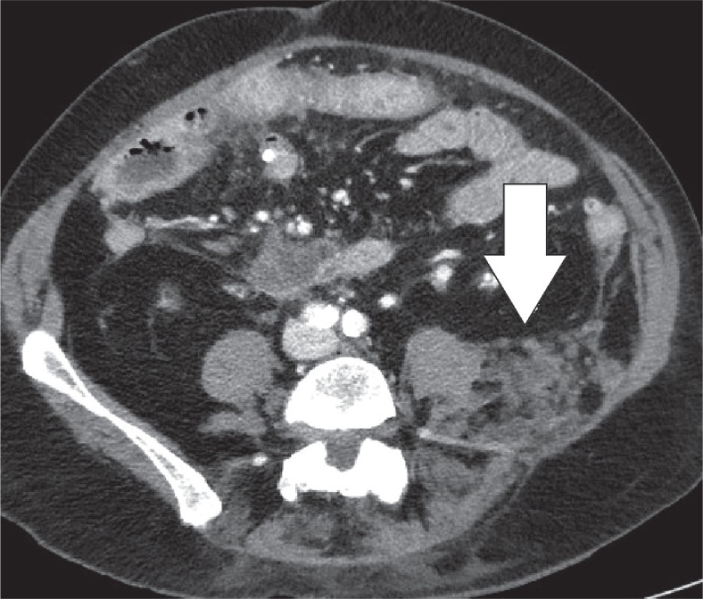
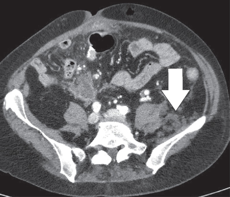
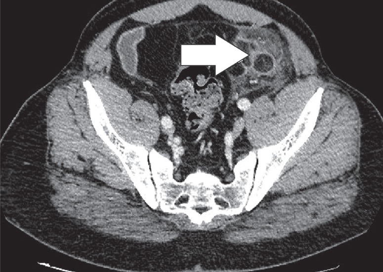
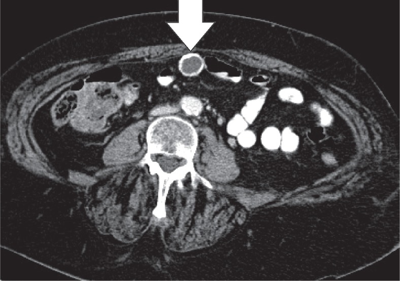
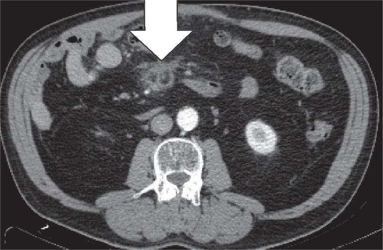
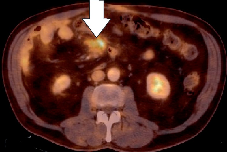
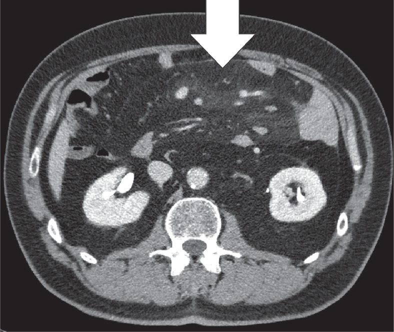
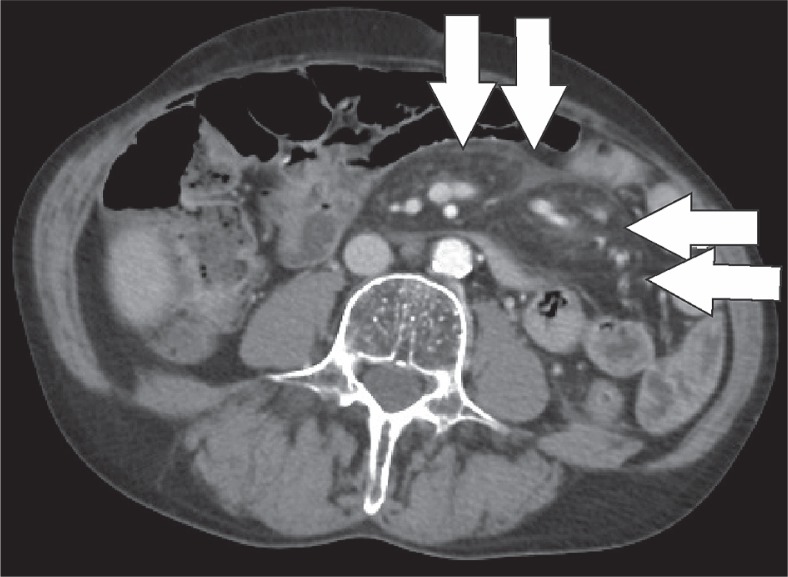
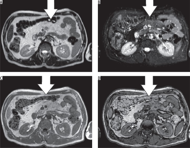
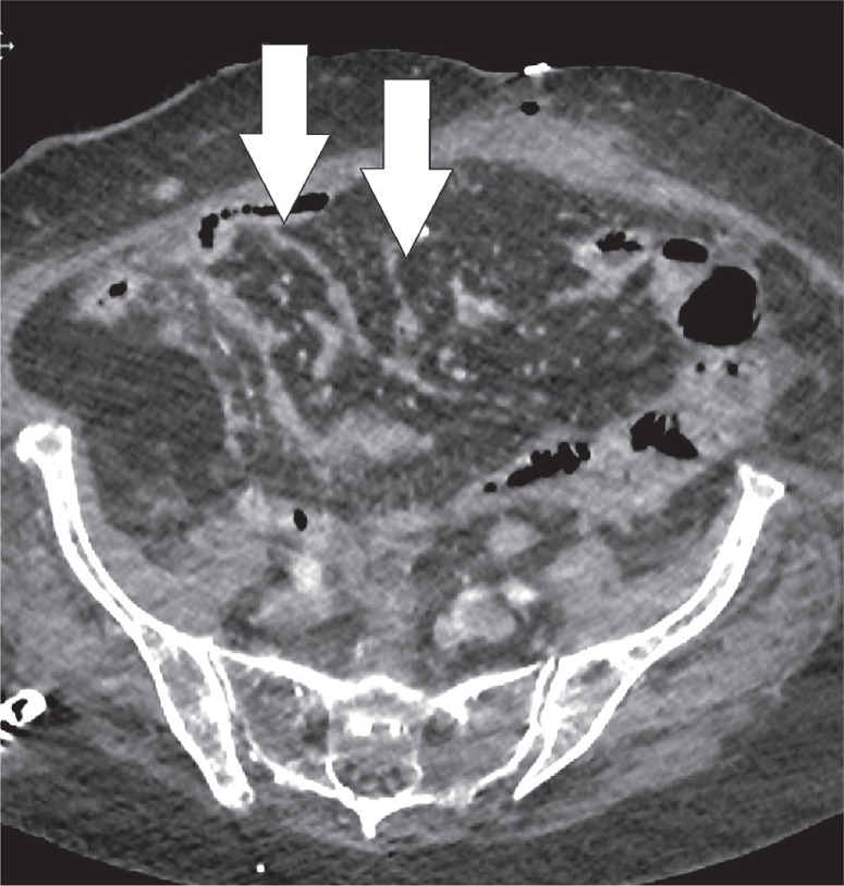
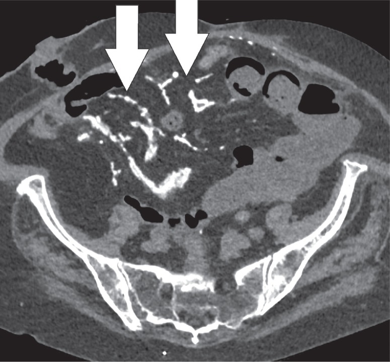
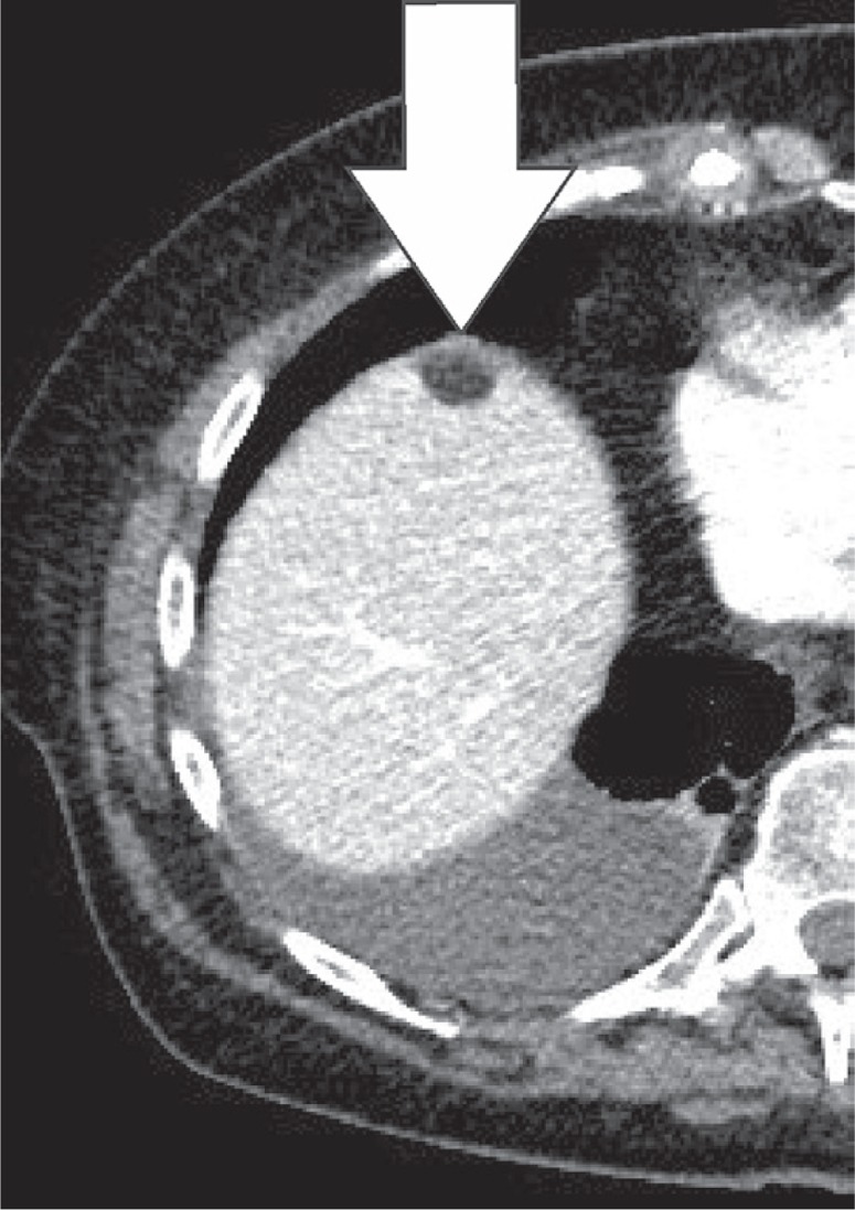
Intra-abdominal fat is abundantly present in both the peritoneum and retroperitoneum. Fat necrosis or inflammation are common findings in abdominal imaging. The most common pathologies that we encounter are epiploic appendagitis, omental infarction, mesenteric panniculitis, and encapsulated fat necrosis. Less common entities that can occur are pancreatic saponification, heterotopic mesenteric ossification, and pseudolipoma of the capsule of Glisson. These entities can mimic more urgent pathologies such as appendicitis, diverticulitis, or malignancies.

摘要

腹腔内脂肪大量存在于腹膜和腹膜后间隙。脂肪坏死或炎症是腹部影像学检查中的常见表现。我们遇到的最常见病理情况是网膜附件炎、网膜梗死、肠系膜脂膜炎和包裹性脂肪坏死。较少见的情况包括胰腺皂化、异位肠系膜骨化和肝门Glisson囊假脂肪瘤。这些病变可能会模仿更紧急的病理情况,如阑尾炎、憩室炎或恶性肿瘤。

https://cdn.ncbi.nlm.nih.gov/pmc/blobs/21fd/7064015/547a24e780e9/PJR-85-39908-g020.jpg
https://cdn.ncbi.nlm.nih.gov/pmc/blobs/21fd/7064015/cd2fe9756598/PJR-85-39908-g001.jpg
https://cdn.ncbi.nlm.nih.gov/pmc/blobs/21fd/7064015/b5ca7f59c77e/PJR-85-39908-g002.jpg
https://cdn.ncbi.nlm.nih.gov/pmc/blobs/21fd/7064015/f7fa1876d65a/PJR-85-39908-g003.jpg
https://cdn.ncbi.nlm.nih.gov/pmc/blobs/21fd/7064015/6c6e980bbf4e/PJR-85-39908-g004.jpg
https://cdn.ncbi.nlm.nih.gov/pmc/blobs/21fd/7064015/43a1d6e21ec6/PJR-85-39908-g005.jpg
https://cdn.ncbi.nlm.nih.gov/pmc/blobs/21fd/7064015/9e768242c198/PJR-85-39908-g006.jpg
https://cdn.ncbi.nlm.nih.gov/pmc/blobs/21fd/7064015/c1ab66651f73/PJR-85-39908-g007.jpg
https://cdn.ncbi.nlm.nih.gov/pmc/blobs/21fd/7064015/502394a05ff4/PJR-85-39908-g008.jpg
https://cdn.ncbi.nlm.nih.gov/pmc/blobs/21fd/7064015/8c60a55141af/PJR-85-39908-g009.jpg
https://cdn.ncbi.nlm.nih.gov/pmc/blobs/21fd/7064015/66b8f0aa011e/PJR-85-39908-g010.jpg
https://cdn.ncbi.nlm.nih.gov/pmc/blobs/21fd/7064015/4a2c6dce0ee6/PJR-85-39908-g013.jpg
https://cdn.ncbi.nlm.nih.gov/pmc/blobs/21fd/7064015/cde7c0315393/PJR-85-39908-g011.jpg
https://cdn.ncbi.nlm.nih.gov/pmc/blobs/21fd/7064015/3ccddd199b98/PJR-85-39908-g012.jpg
https://cdn.ncbi.nlm.nih.gov/pmc/blobs/21fd/7064015/09e49410955e/PJR-85-39908-g014.jpg
https://cdn.ncbi.nlm.nih.gov/pmc/blobs/21fd/7064015/7821313a8caf/PJR-85-39908-g015.jpg
https://cdn.ncbi.nlm.nih.gov/pmc/blobs/21fd/7064015/37d211ee5e9a/PJR-85-39908-g016.jpg
https://cdn.ncbi.nlm.nih.gov/pmc/blobs/21fd/7064015/6248b0896fe3/PJR-85-39908-g017.jpg
https://cdn.ncbi.nlm.nih.gov/pmc/blobs/21fd/7064015/4f16a142f55b/PJR-85-39908-g018.jpg
https://cdn.ncbi.nlm.nih.gov/pmc/blobs/21fd/7064015/6a720de8f2f4/PJR-85-39908-g019.jpg
https://cdn.ncbi.nlm.nih.gov/pmc/blobs/21fd/7064015/547a24e780e9/PJR-85-39908-g020.jpg

相似文献

1
Fat misbehaving in the abdominal cavity: a pictorial essay.腹腔内的脂肪异常:一篇图文并茂的文章。
Pol J Radiol. 2020 Jan 21;85:e32-e38. doi: 10.5114/pjr.2020.93070. eCollection 2020.
2
Imaging of Mischievous Intra-abdominal Fat Presenting with Abdominal Pain: A Pictorial Review.表现为腹痛的腹腔内有害脂肪的影像学:图文综述
Euroasian J Hepatogastroenterol. 2022 Jan-Jun;12(1):45-49. doi: 10.5005/jp-journals-10018-1355.
3
Acute epiploic appendagitis and its mimics.急性网膜附件炎及其模仿疾病。
Radiographics. 2005 Nov-Dec;25(6):1521-34. doi: 10.1148/rg.256055030.
4
Omental infarction and its mimics: imaging features of acute abdominal conditions presenting with fat stranding greater than the degree of bowel wall thickening.网膜梗死及其类似病变:以脂肪浸润程度大于肠壁增厚程度为表现的急性腹部疾病的影像学特征。
Emerg Radiol. 2015 Aug;22(4):431-6. doi: 10.1007/s10140-015-1302-0. Epub 2015 Mar 1.
5
[Intraabdominal fat necrosis].[腹腔内脂肪坏死]
Radiologia. 2012 Sep-Oct;54(5):449-56. doi: 10.1016/j.rx.2011.07.006. Epub 2011 Oct 22.
6
Epiploic appendagitis and omental infarction: pitfalls and look-alikes.网膜附件炎和网膜梗死:陷阱与相似病症
Abdom Imaging. 2002 Jan-Feb;27(1):20-8. doi: 10.1007/s00261-001-0056-1.
7
Epiploic appendagitis: A rare cause of acute abdomen.网膜附件炎:急性腹痛的罕见病因。
Radiol Case Rep. 2018 Mar 23;13(3):599-601. doi: 10.1016/j.radcr.2018.02.022. eCollection 2018 Jun.
8
Evaluating the Patient with Right Lower Quadrant Pain.评估右下象限疼痛的患者。
Radiol Clin North Am. 2015 Nov;53(6):1159-70. doi: 10.1016/j.rcl.2015.06.004. Epub 2015 Aug 13.
9
Intraabdominal focal fat infarction in a 75-year-old woman presenting as acute abdomen.75 岁女性,以急腹症为表现的腹腔局灶性脂肪梗死。
Am J Emerg Med. 2012 Nov;30(9):2096.e3-5. doi: 10.1016/j.ajem.2012.01.012. Epub 2012 Mar 16.
10
Know the name: acute epiploic appendagitis-CT findings and review of literature.了解名称:急性网膜附件炎——CT表现及文献综述
AME Case Rep. 2018 Mar 7;2:8. doi: 10.21037/acr.2018.02.02. eCollection 2018.

引用本文的文献

1
Endometriosis in Patients Undergoing Plastic Surgical Procedures: A Case Report and Review of the Literature.接受整形手术患者的子宫内膜异位症:一例报告及文献综述
Plast Reconstr Surg Glob Open. 2024 Jun 17;12(6):e5904. doi: 10.1097/GOX.0000000000005904. eCollection 2024 Jun.
2
Focal necrosis of the falciform ligament as a rare cause of abdominal pain: A case report.镰状韧带局灶性坏死作为腹痛的罕见原因:一例报告
SAGE Open Med Case Rep. 2024 May 15;12:2050313X241252738. doi: 10.1177/2050313X241252738. eCollection 2024.
3
Characterization of hypermetabolic lymph nodes after SARS-CoV-2 vaccination using PET-CT derived node-RADS, in patients with melanoma.

本文引用的文献

1
Heterotopic mesenteric and abdominal wall ossification - Two case reports in one institution.异位肠系膜和腹壁骨化——同一机构的两例病例报告
Int J Surg Case Rep. 2017;37:22-25. doi: 10.1016/j.ijscr.2017.06.004. Epub 2017 Jun 8.
2
Post-pancreatitis Fat Necrosis Mimicking Carcinomatosis.胰腺炎后脂肪坏死酷似癌性腹膜炎。
Radiol Case Rep. 2015 Dec 7;3(2):192. doi: 10.2484/rcr.v3i2.192. eCollection 2008.
3
Peritoneal lymphomatosis: CT and PET/CT findings and how to differentiate between carcinomatosis and sarcomatosis.腹膜淋巴瘤病:CT 和 PET/CT 的表现,以及如何鉴别癌性腹膜转移和肉瘤腹膜转移。
基于 PET-CT 构建的淋巴结放射学报告数据系统(node-RADS)对 SARS-CoV-2 疫苗接种后高代谢性淋巴结的特征分析,应用于黑色素瘤患者。
Sci Rep. 2023 Oct 26;13(1):18357. doi: 10.1038/s41598-023-44215-2.
4
Epiploic Adipose Tissue (EPAT) in Obese Individuals Promotes Colonic Tumorigenesis: A Novel Model for EPAT-Dependent Colorectal Cancer Progression.肥胖个体的网膜脂肪组织(EPAT)促进结肠肿瘤发生:一种依赖EPAT的结直肠癌进展新模型。
Cancers (Basel). 2023 Feb 3;15(3):977. doi: 10.3390/cancers15030977.
5
Retroperitoneal Fat Necrosis in Response to an Episode of Acute Pancreatitis.急性胰腺炎发作后出现的腹膜后脂肪坏死
Case Rep Gastrointest Med. 2021 Sep 23;2021:1051077. doi: 10.1155/2021/1051077. eCollection 2021.
6
Omental Infarction in a Child-Conservative Management as an Effective and Safe Strategy in Diagnosis and Treatment.小儿脐肠系膜梗死:一种有效的诊断和治疗策略
Int J Environ Res Public Health. 2021 Jul 29;18(15):8057. doi: 10.3390/ijerph18158057.
7
Not only fat: omental infarction and its mimics in children. Clinical and ultrasound findings: a pictorial review.不仅是脂肪:儿童的网膜梗死及其模拟疾病。临床和超声表现:图像综述。
J Ultrasound. 2020 Dec;23(4):621-629. doi: 10.1007/s40477-020-00492-5. Epub 2020 Jul 4.
Cancer Imaging. 2013 Apr 15;13(2):162-70. doi: 10.1102/1470-7330.2013.0018.
4
The "misty mesentery": mesenteric panniculitis and its mimics.
AJR Am J Roentgenol. 2013 Feb;200(2):W116-23. doi: 10.2214/AJR.12.8493.
5
Image of the month. Encapsulated fat necrosis.
Arch Surg. 2011 Dec;146(12):1447-8. doi: 10.1001/archsurg.146.12.1447.
6
Imaging manifestations of abdominal fat necrosis and its mimics.腹部脂肪坏死及其类似物的影像学表现。
Radiographics. 2011 Nov-Dec;31(7):2021-34. doi: 10.1148/rg.317115046.
7
Release of inflammatory mediators by adipose tissue during acute pancreatitis.急性胰腺炎时脂肪组织释放炎症介质。
J Pathol. 2010 Jun;221(2):175-82. doi: 10.1002/path.2691.
8
Benign hepatic tumors and iatrogenic pseudotumors.良性肝肿瘤和医源性假瘤。
Radiographics. 2009 Jan-Feb;29(1):211-29. doi: 10.1148/rg.291085099.
9
Is it from the mesentery or the omentum? MDCT features of various pathologic conditions in intraperitoneal fat planes.它来自肠系膜还是大网膜?腹腔脂肪平面各种病理状况的MDCT特征。
Surg Radiol Anat. 2009 Jan;31(1):3-11. doi: 10.1007/s00276-008-0381-y. Epub 2008 Jul 4.
10
Heterotopic ossification after blunt abdominal trauma.钝性腹部创伤后异位骨化
J Trauma. 2008 Dec;65(6):1536-9. doi: 10.1097/01.ta.0000228884.03835.d9.